
Endoscopic apparatus and method

a technology of endoscope and endoscope tube, which is applied in the field of endoscope devices, can solve the problems of limiting the size of the distal assembly on the distal end of the endoscope, limiting the size of tissue samples obtained, and preventing the use of forceps

Inactive Publication Date: 2007-01-30
SCI MED LIFE SYST
View PDF19 Cites 58 Cited by
  • Summary
  • Abstract
  • Description
  • Claims
  • Application Information

AI Technical Summary

Benefits of technology

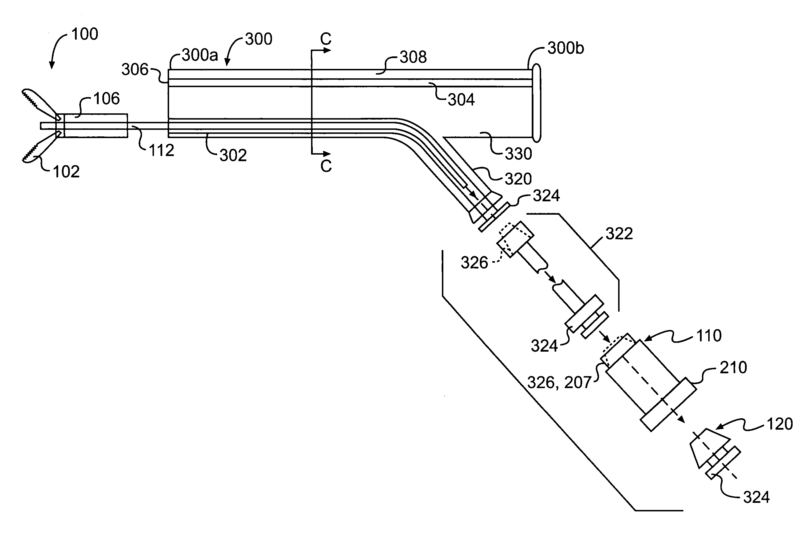
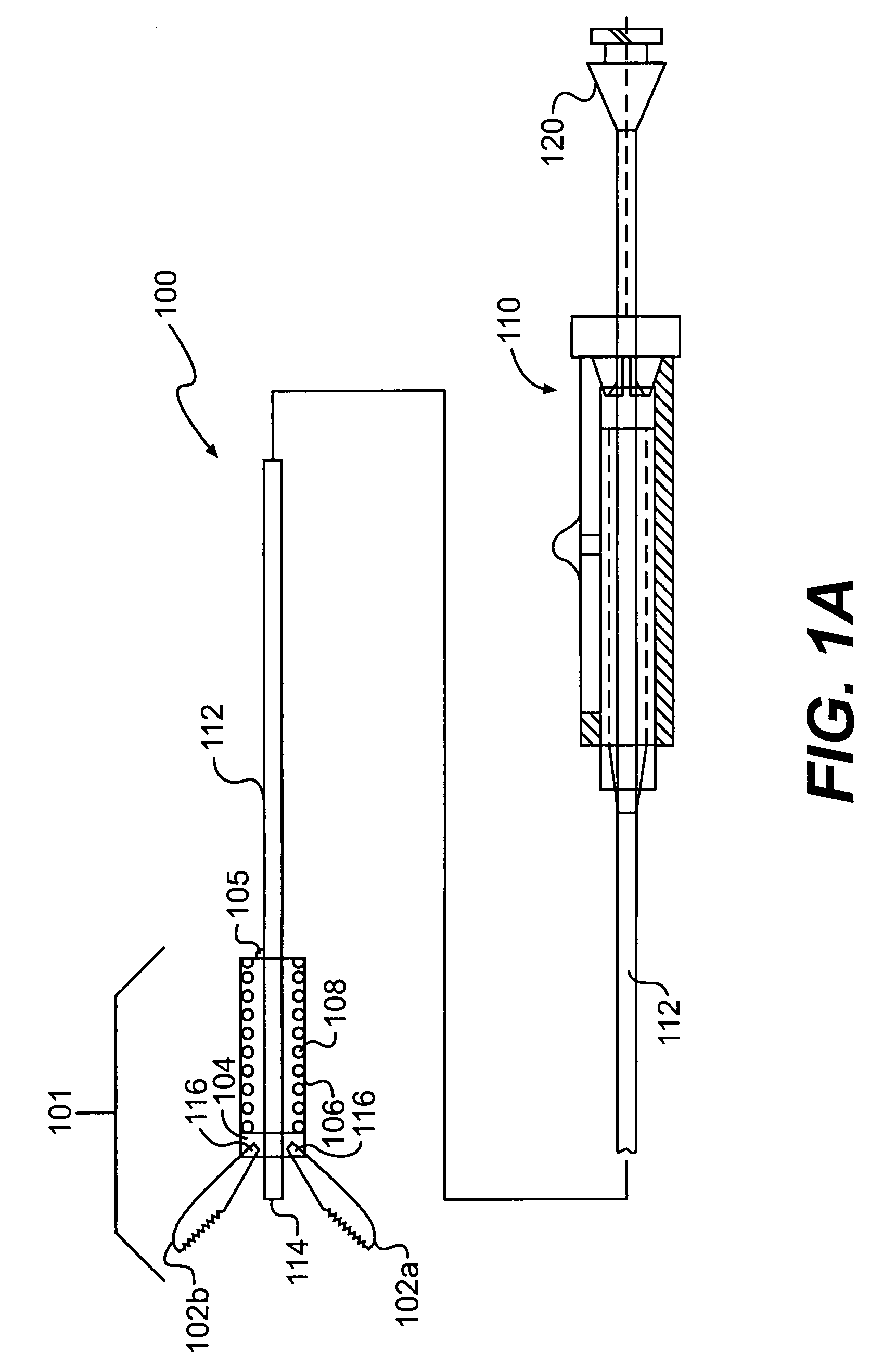
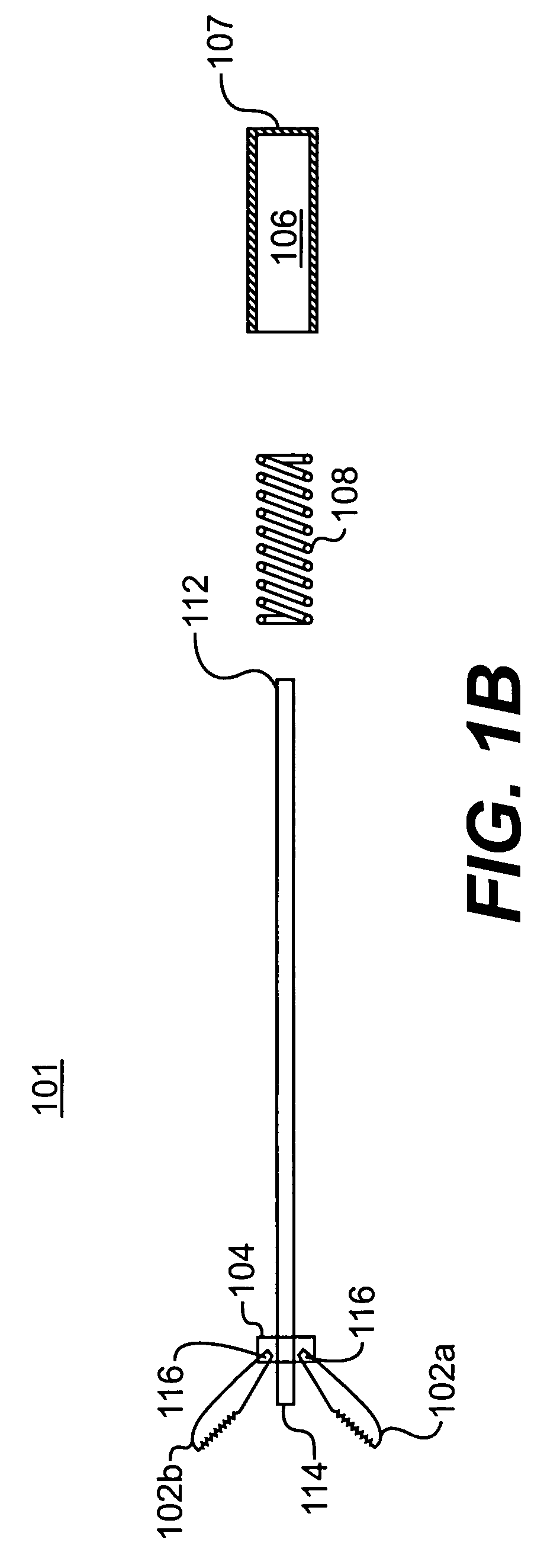
"The present invention is a medical device that can be inserted into an endoscope to perform procedures. It has a large distal assembly that is designed to fit properly and can be loaded into the endoscope before insertion. The distal assembly has a closed configuration with a profile larger than the diameter of the endoscope channel, making it suitable for use in the body. The device can be activated to perform the desired procedure by opening and closing the distal assembly. The technical effects of this invention include improved access and visualization for procedures performed with an endoscope."

Problems solved by technology

The size of a distal assembly on the distal end of the endoscopic device, such as forceps, stent delivery devices, biopsy needles, and electrocoagulation probes, is limited by the diameter of the endoscope's working channel.
The jaw members must be small enough to fit through the working channel of the endoscope, however, which limits the size of the tissue samples obtained.
The size limitation of the jaws also prohibits the use of forceps for other applications, such as grasping gall stones and kidney stones or removing or displacing larger size tissue samples.
A working channel of an endoscope that is sized to accommodate an endoscopic device with a larger profile or diameter can inhibit both the flexibility of the endoscope and / or the ability of the endoscope to house other functional components, such as visualization means, or may increase the outer diameter of the endoscope to an unusable size.

Method used


Image

  • Endoscopic apparatus and method
  • Endoscopic apparatus and method
  • Endoscopic apparatus and method

Examples


Embodiment Construction

[0001]1. Field of the Invention

[0002]The present invention relates generally to endoscopic devices, including those used for obtaining biopsies or for performing other medical procedures. More particularly, the present invention relates to an endoscopic device having a suitably large distal assembly to properly perform its intended function.

[0003]2. Background of the Invention

[0004]Endoscopic devices for use in medical procedures typically are passed through a working channel of an endoscope positioned in a body cavity in order to reach an operative site at a distal end of the endoscope. For purposes of this description, “distal” refers to the end extending into a body and “proximal” refers to the end extending out of the body. The size of a distal assembly on the distal end of the endoscopic device, such as forceps, stent delivery devices, biopsy needles, and electrocoagulation probes, is limited by the diameter of the endoscope's working channel. Because the endoscopic device, suc...


PUM


Abstract

The present invention is directed to a medical device, such as an endoscopic device, configured to be loaded into a channel of an endoscope prior to insertion of the endoscope into a body, and a method of performing an operative procedure with an endoscopic device. The endoscopic device comprises an elongate member for insertion into the channel of the endoscope, wherein a length of the elongate member is greater than a length of the channel of the endoscope. The endoscopic device also comprises a distal assembly connected to a distal portion of the elongate member and operable to perform an endoscopic operation, wherein the distal assembly has an open configuration and a closed configuration with a profile larger than a diameter of the channel of the endoscope, wherein the distal assembly is adapted to be exterior to the channel when the endoscope is inserted into the body.

Description

DESCRIPTION OF THE INVENTION[0001]1. Field of the Invention[0002]The present invention relates generally to endoscopic devices, including those used for obtaining biopsies or for performing other medical procedures. More particularly, the present invention relates to an endoscopic device having a suitably large distal assembly to properly perform its intended function.[0003]2. Background of the Invention[0004]Endoscopic devices for use in medical procedures typically are passed through a working channel of an endoscope positioned in a body cavity in order to reach an operative site at a distal end of the endoscope. For purposes of this description, “distal” refers to the end extending into a body and “proximal” refers to the end extending out of the body. The size of a distal assembly on the distal end of the endoscopic device, such as forceps, stent delivery devices, biopsy needles, and electrocoagulation probes, is limited by the diameter of the endoscope's working channel. Becaus...

Claims


Application Information

Patent Timeline
Patent Type & Authority Patents(United States)
IPC IPC(8): A61B17/28
CPCA61B17/29A61B2017/2901A61B2017/2931A61B2017/2929A61B2017/2905
Inventor CHU, MICHAEL S. H.
Owner SCI MED LIFE SYST
Learn More