

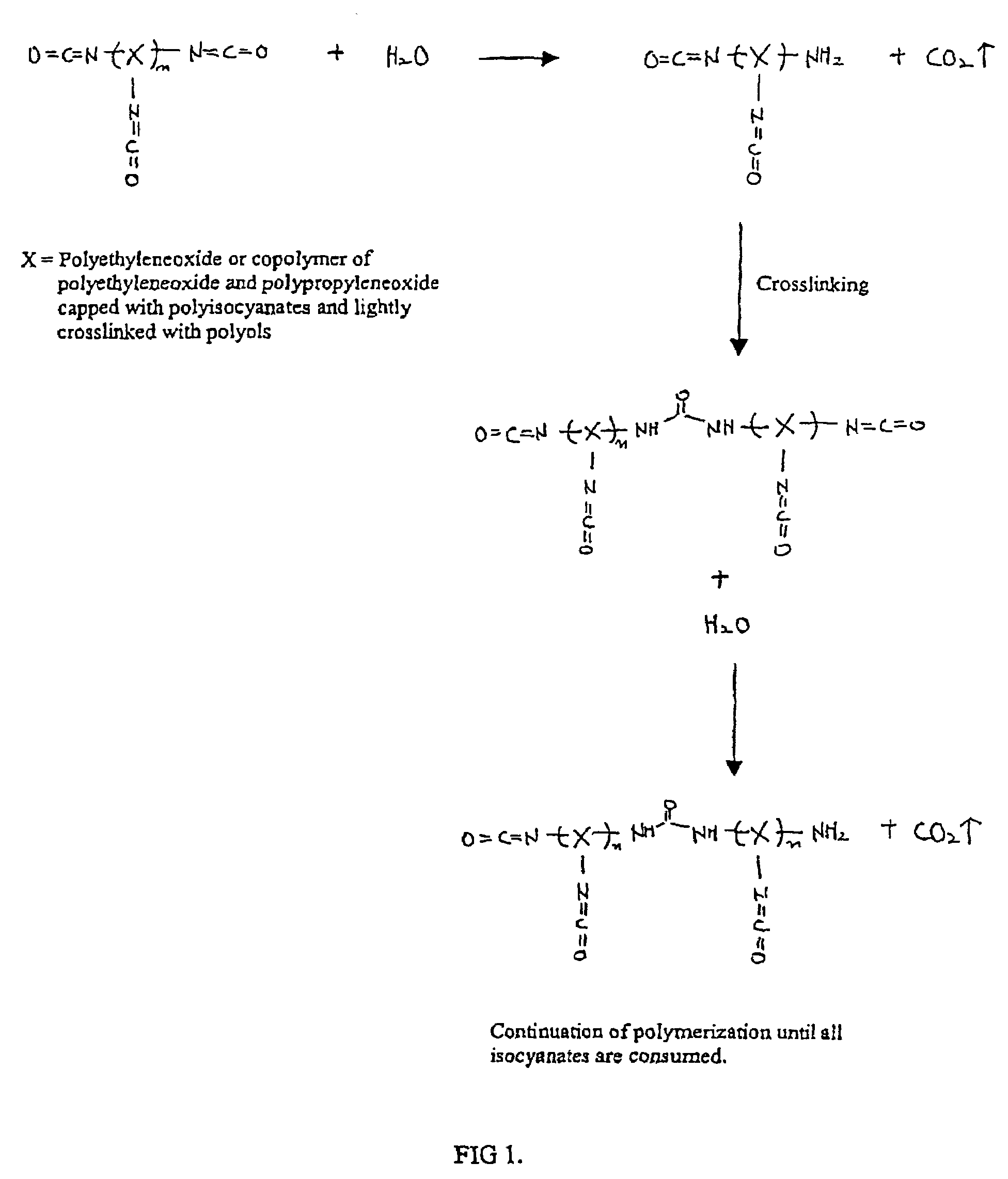
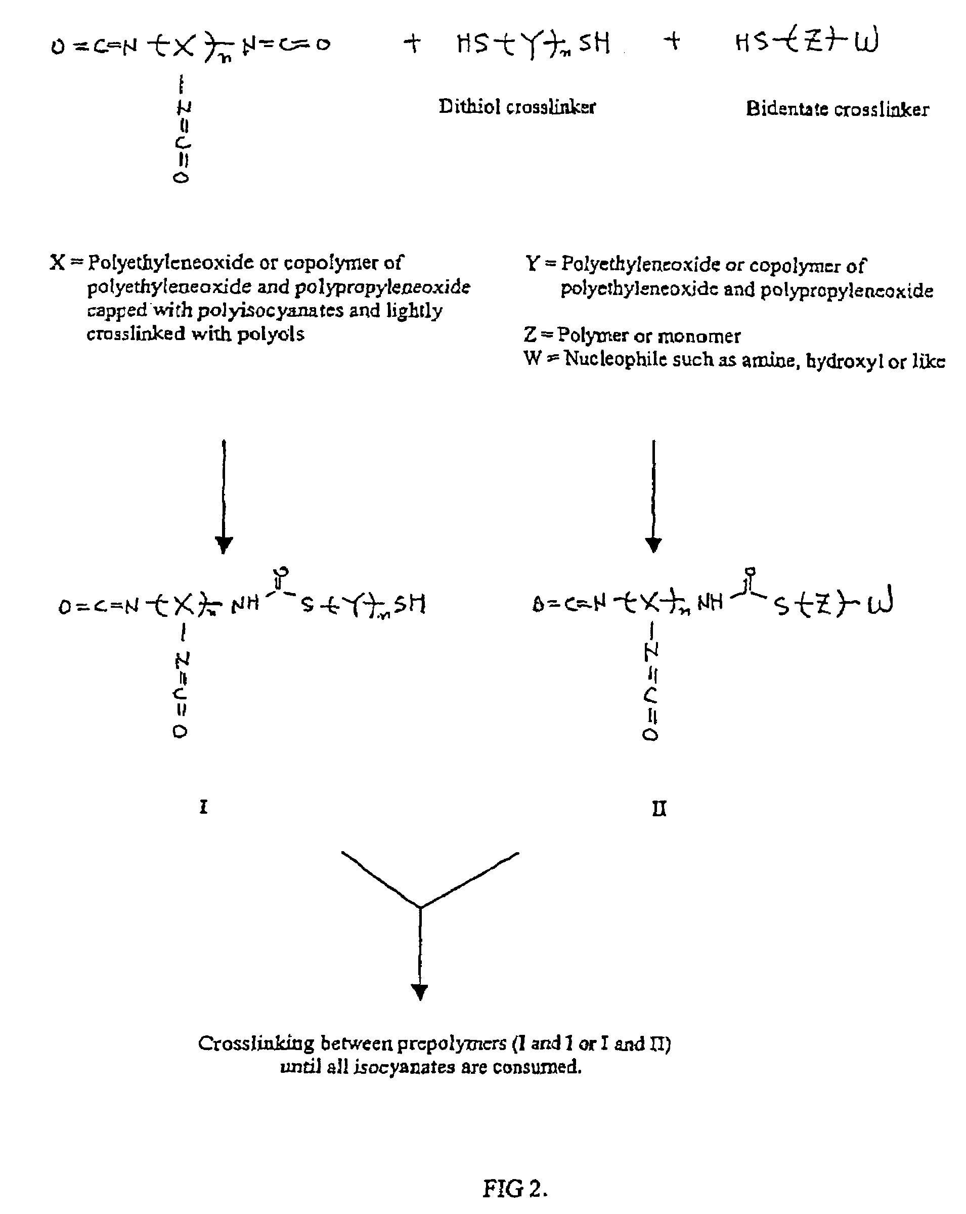
Methods and gel compositions for encapsulating living cells and organic molecules
a technology of organic molecules and gel compositions, applied in the field of systems and methods for forming polyurethane hydrogels, can solve the problem that the polymerization process is truly biocompatible, and achieve the effects of high throughput biological testing, simple and easy process development, and potential employment of such materials
- Summary
- Abstract
- Description
- Claims
- Application Information
AI Technical Summary
Benefits of technology
Problems solved by technology
Method used
Image

Examples
example 1
[0036]Solution A was prepared by mixing 0.075 g of Hypol PreMa G-50 (Hampshire Chemical Corp.) and 1.5 mL of 50 mM aqueous phosphate buffer, at pH 7.0 with 80 mM sodium chloride. Solution B was prepared by dissolving 30 mg of PEG-(thiol)2 (mw=3,400) and 2 mg of free base cysteine (Sigma Chemical Co.) (mw=121) in 1 mL of 50 mM phosphate buffer, at pH 7.0 with 60 mM sodium chloride. Solution C was prepared by mixing 100 μL of Solution B with 10 μL of goat lymphocytes in Dulbecco's phosphate-buffered saline. Finally, 200 μL of Solution A was mixed with 50 μL of Solution C, and the resulting solution was microspotted onto amine-treated glass (Silanated Slides, Cell Associates, Inc.) with the use of 5 microliter glass microcapillary tubes. The hydrogel spots were polymerized carefully in a humidity box, at about 95% RH, to avoid dehydration. This formulation polymerized within 5–10 minutes. After polymerization, the hydrogel spots were physically stable and strongly attached to the glass...
example 2
[0037]Solution A was prepared by mixing 0.1 g of Hypol PreMa G-50 (Hampshire Chemical Corp.) and 1 mL of 50 mM phosphate buffer, at pH 7.0 with 80 mM sodium chloride. Solution B was prepared by the same procedure as in Example 1. Solution C was prepared by mixing 40 μL of Solution B with 70 μL of goat lymphocytes in Dulbecco's phosphate-buffered saline. Finally, 100μL of Solution A was mixed with 100 μL of Solution C, and the resulting solution was microspotted using the same procedure as in Example 1. The formulation polymerized in 5 minutes, and the hydrogel spots were treated with Dulbecco's modified phosphate-buffered saline solution and incubated at 37° C. for 1 day to 3 days in RPM1640 cell media. The viability of lymphocytes was examined with a light microscope using AlamarBlue which demonstrated viable encapsulated cells.
example 3
[0038]Solution A was prepared by mixing 0.1 g of Hypol PreMa G-50 (Hampshire Chemical Corp.) and 1 mL of 50 mM phosphate buffer, at pH 7.0 with 80 mM sodium chloride. Solution B was prepared by the same procedure as in Example 1. Solution C was prepared by mixing 40 μL of Solution B with 70 μL of E. coli in Dulbecco's phosphate-buffered saline. Finally, 100 μL of Solution A was mixed with 100 μL of Solution C, and the resulting solution was placed into a disposable culture tube. The formulation polymerized in 5 minutes, and the hydrogel was treated with Dulbecco's modified phosphate-buffered saline solution and incubated at 37° C. for 1 day in RPM1640 cell media. Viability and growth of E. coli were confirmed by observing turbidity in the hydrogel after 1 day.
PUM
| Property | Measurement | Unit |
|---|---|---|
| pKa | aaaaa | aaaaa |
| pKa | aaaaa | aaaaa |
| pH | aaaaa | aaaaa |
Abstract
Description
Claims
Application Information