
Device for use in eye surgery

a technology for eye surgery and sealing, applied in the field of sealing devices, can solve the problems of permanent optical problems, unable to meet the needs of patients, and liquid material can leak through the capsulorhexis, and achieve the effect of convenient removal and large area

Inactive Publication Date: 2007-02-27
AMO GRONINGEN
View PDF10 Cites 60 Cited by
  • Summary
  • Abstract
  • Description
  • Claims
  • Application Information

AI Technical Summary

Benefits of technology

The invention is a sealing device for the capsulorhexis of the eye that prevents leakage of liquid or gas through the capsulorhexis after a lens implantation process. The device consists of a plug part made of a deformable polymer that can be introduced into the capsular bag through a small incision and adjusted in position by a removable adjusting means. The plug part is designed to leave no permanent parts outside the capsular bag. The device can compensate for refractive errors and wavefront aberrations of the eye. It can be easily controlled and removed after the lens forming process is completed. The sealing device helps to improve the stability and accuracy of the lens implantation process.

Problems solved by technology

It has been identified as a problem in capsular bag filling processes that during the injection and before the final lens is formed liquid material can leak through the capsulorhexis.
A problem with this plug is that it is clamped and / or glued in the rhexis.
Since the plug is relatively large and adapted to stay in the rhexis permanent optical problems could arise.
Furthermore, it needs a relatively large opening in the eye to be implanted.
The tube could cause a leak.
Also this plug suffers from the drawback that it permanently locates parts of its structure outside the capsular bag after finalizing the surgery.
Consequently, the protruding parts may risk to compromise surrounding delicate eye tissues, including the iris, while they also risk generating unwanted optical side effects.

Method used


Image

  • Device for use in eye surgery
  • Device for use in eye surgery
  • Device for use in eye surgery

Examples


first embodiment

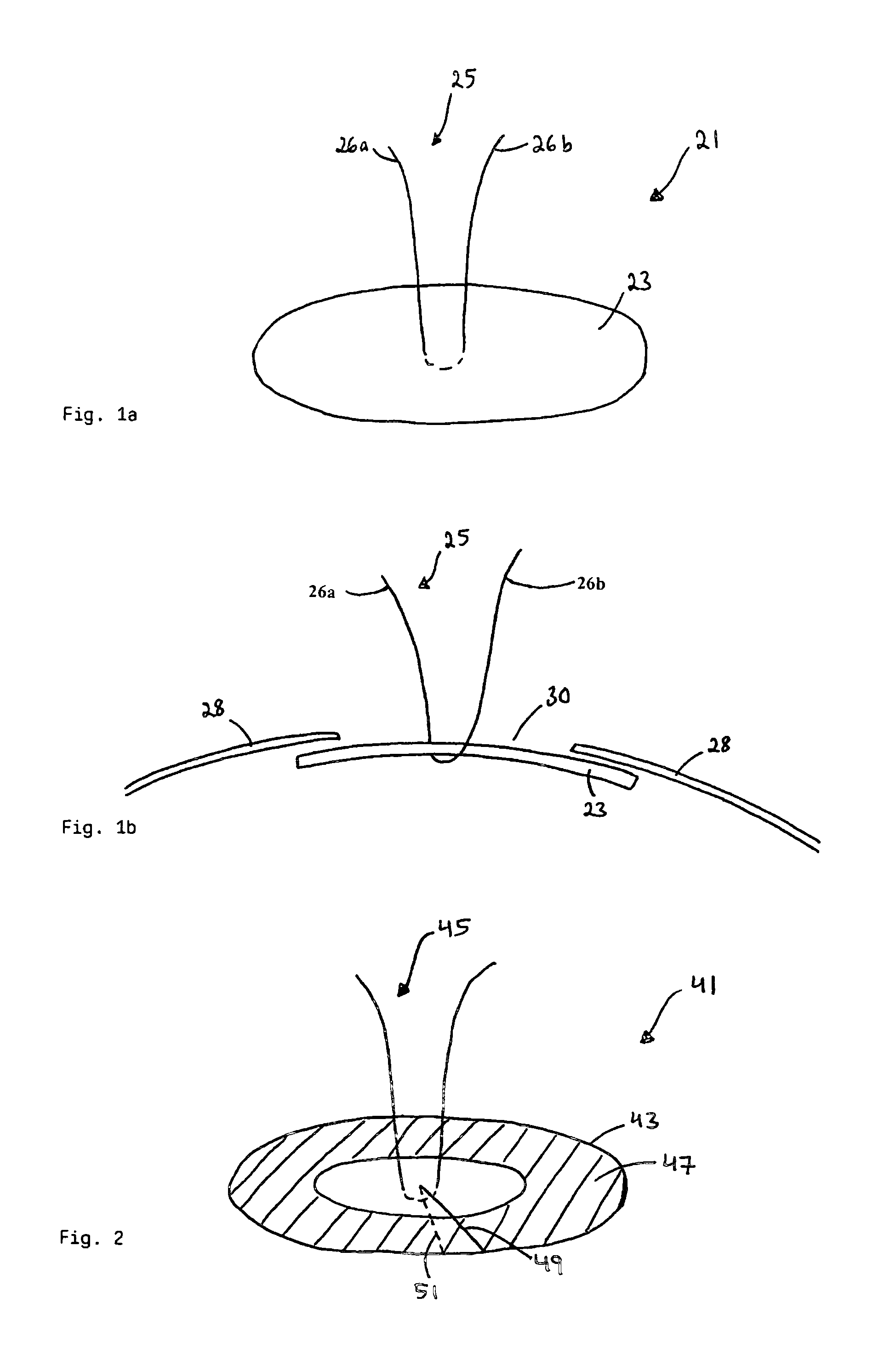
[0027]FIG. 1a is a schematic view from above of a sealing device according to the invention.

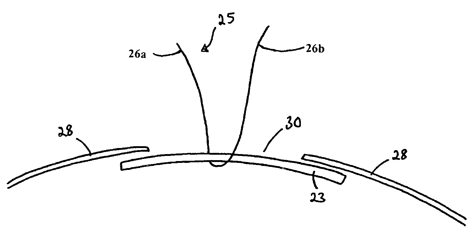
[0028]FIG. 1b is a side view of the sealing device in FIG. 1a inserted into a capsular bag.

second embodiment

[0029]FIG. 2 is a view from above of the sealing device according to the invention.

third embodiment

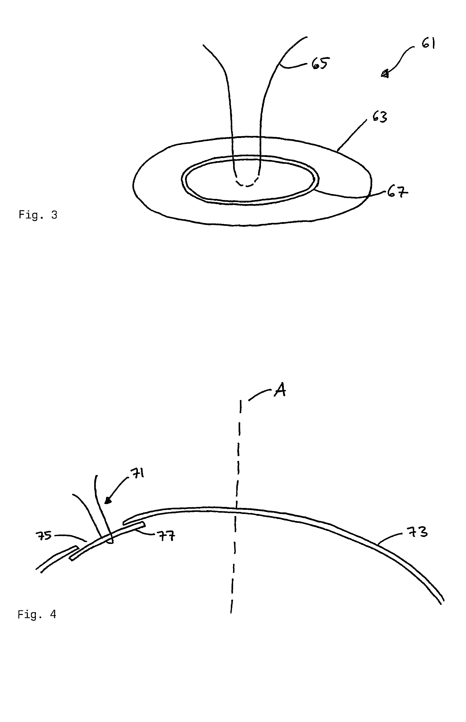
[0030]FIG. 3 is a view from above of the sealing device according to the invention.

[0031]FIG. 4 is a side view of a sealing device according to the invention inserted into a capsular bag.


PUM


Abstract

A method of manufacturing an intraocular lens inside a capsular bag after the natural lens has been removed employs a sealing device comprising a plug part adapted to seal a rhexis in a capsular bag, thus preventing displacement through the rhexis of a lens-forming liquid material injected through the rhexis and adapted to replace the natural lens and form an intraocular lens implant. The plug part has a slightly larger area than the capsulorhexis and is made of a deformable polymer. The sealing device further comprises an adjusting means connected to the plug part and adapted to position the plug part to a desired location.

Description

FIELD OF THE INVENTION[0001]The present invention relates to a sealing device comprising a plug part adapted to seal a capsulorhexis of a capsular bag used in surgical processes involving insertion of lens-forming liquid to replace the natural lens and forming an intraocular lens implant. It also relates to a method providing visual correction including manufacturing an intraocular lens inside a capsular bag after that the natural lens has been removed.BACKGROUND OF THE INVENTION[0002]A technique for removing a catarcteous and / or presbyopic natural lens from the capsular bag of the eye and replacing it by a lens-forming liquid material injected directly into the capsular bag is under development. This is described in for example the patent application with application number SE 0001934-9. The liquid material is a partially polymerized material, which can undergo a curing process in the eye and thereby form a solid lens implant. The lens implant acts as a substitute for the natural l...

Claims


Application Information

Patent Timeline
Patent Type & Authority Patents(United States)
IPC IPC(8): A61F2/16A61F9/007
CPCA61F2/1616A61F9/007A61F2/1662
Inventor TERWEE, THOMASKOOPMANS, STEVEN
Owner AMO GRONINGEN
Learn More