Method and apparatus for determining the cerebral state of a patient with fast response

a technology fast response, which is applied in the field of method and apparatus for can solve the problems of excessive deep anesthesia, unfavorable patient recovery, and inability to accurately determine etc., and achieve the effect of accurately determining the cerebral state of a patien

Inactive Publication Date: 2007-06-05
GE HEALTHCARE FINLAND
View PDF30 Cites 39 Cited by
  • Summary
  • Abstract
  • Description
  • Claims
  • Application Information

AI Technical Summary

Benefits of technology

[0041]An object of the present invention is to provide an improved method and apparatus for accurately determining the cerebral state of a patient, including the hypnotic or consciousness state of a patient and the depth of anesthesia that a patient is experiencing.

Problems solved by technology

If the anesthesia is not sufficiently deep, the patient may maintain or gain consciousness during a surgery, or other medical procedure, resulting in an extremely traumatic experience for the patient, anesthesiologist, and surgeon.
On the other hand, excessively deep anesthesia reflects an unnecessary consumption of anesthetic agents, most of which are expensive.
A second reason for the continuing study and attention being given to monitoring the hypnotic condition of a patient arises because of its difficulty: that is, anesthetic agents alter the activity and state of the patient's brain and these changes are not always easy to detect.
While useful for research and other purposes, an OAAS scale provides only a limited number of scaling levels and is limited in practical use because of the attention required from the anesthesiologist and the use of painful stimuli.
However, this gross characterization is too imprecise and unreliable to use as an indication of such a critical medical aspect as the extent of hypnosis.
Further, EEG signal changes during anesthesia may not fully correlate with changes in the hypnotic state of the patient.
This means that it is not possible to exactly predict future values of the EEG from past values in the manner that, for example, the shapes of past QRS complexes in an ECG signal can be used to predict future values for analytical and diagnostic purposes.
Time-domain based EEG analysis methods have not proven greatly successful in clinical applications since the results do not behave in a completely consistent manner.
However, because the calculation must be performed using complex number arithmetic for several thousand f1, f2, and f1+f2 frequency combinations, the computations to obtain bispectral information are rather arduous.
While parameters of the foregoing types can detect changes in the EEG caused by anesthetic agents and hence are useful in determining the depth of anesthesia, they suffer from an inability to be calibrated to behavioral endpoints and because of their sensitivity to the different EEG patterns induced by different anesthetic agents.
The bispectral information in the SyncFastSlow subparameter does not, by itself, give sufficient information over the range of hypnosis thus requiring combination with the other subparameters.
Thus, this index may be not sufficiently fast to detect changes in the state of a patient as is required in the clinical situation.
This can be a serious problem in the use of the BIS index.
However, the lessened amount of anesthetic agent increases the risk that the patient will awaken during surgery.

Method used


Image

  • Method and apparatus for determining the cerebral state of a patient with fast response
  • Method and apparatus for determining the cerebral state of a patient with fast response
  • Method and apparatus for determining the cerebral state of a patient with fast response

Examples


first embodiment

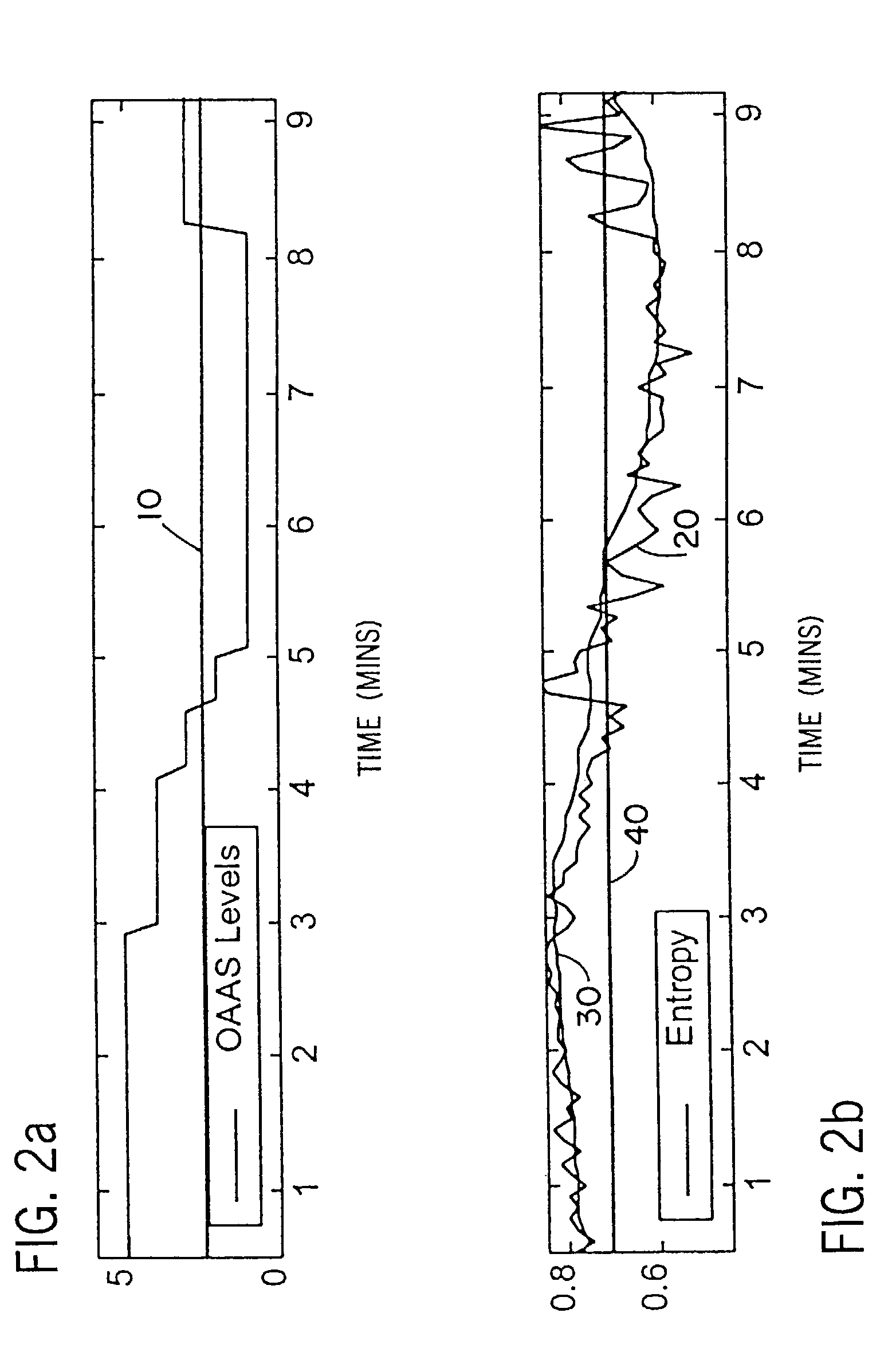
[0097]In providing an entropy based indicator obtained from a frequency range containing both EEG and EMG spectra, it is to be appreciated that this represents a departure from the customary expression of, particularly, EMG signal data. That is, as noted above in connection with the present invention, EMG signal characteristics are customarily expressed as a voltage amplitude, for example as a root mean squared spectral amplitude, and the amplitude of the voltage will vary as a result of variations in the EMG signal data. By contrast, EEG entropy is a dimensionless quantity which describes the amount of disorder in the signal. When viewed from the entropic standpoint, the effect of EMG activity on the EEG signal is to create high frequency noise which increases the entropy of the combined signal. Entropy varies from 0 to 1, and the values are independent of the amplitude of the signal. The entropic expression of EEG signal data and the amplitude expression of the EMG signal data thu...

second embodiment

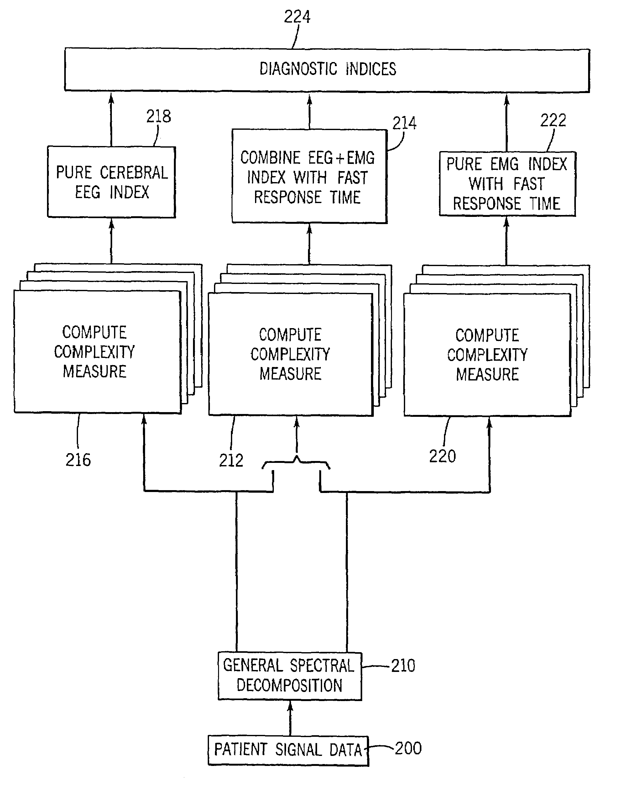
[0099]the invention is explained using a frequency range of 0.5 Hz to about 150 Hz. It is deemed preferable to divide the extended frequency range into three-bands: a 0.5–47 Hz band; a 63–97 Hz band; and a 123–147 Hz band. The range is divided into the three bands at these frequencies in order to avoid the power line harmonics at 50 / 100 Hz or 60 / 120 Hz depending on the frequency of the alternating current power mains. The lowest band contains most of the EEG components, while the two upper bands include primarily EMG activity.

[0100]FIG. 8 is a flow chart showing the steps of producing an improved diagnostic indication or index using a widened frequency range for the computation of spectral entropy in accordance with the second embodiment of the present invention. In step 200, the signal data corresponding to the biopotential signals appearing in the electrodes placed on the scalp of the patient is obtained. In step 210, the signal data is subjected to spectral decomposition, as by u...


PUM


Abstract

A method and apparatus for ascertaining the cerebral state of a patient. The method/apparatus may find use in ascertaining the depth of anesthesia of the patient. In one embodiment, the entropy of the patient's EEG signal data is determined as an indication of the cerebral state. A frequency domain power spectrum quantity is obtained from the patient's EMG signal data. The latter quantity can be updated more frequently than the EEG entropy due to its higher frequency. The EEG entropy indication and the EMG power spectrum indication can be combined into a composite indicator that provides an immediate indication of changes in the cerebral state of the patient. In another embodiment, the frequency range over which the entropy of the biopotential signal from the patient is determined is broadened to encompass both EEG signal data and EMG signal data and the entropy so determined used as an indication of the patient's cerebral state.

Description

CROSS-REFERENCE TO RELATED APPLICATIONS[0001]The present application is a divisional application of U.S. patent application Ser. No. 09 / 688,891, filed Oct. 16, 2000, and now U.S. Pat. No. 6,731,975.BACKGROUND OF THE INVENTION[0002]The present invention relates to a method and apparatus for determining the cerebral state of a patient. One application of the method and apparatus is determining the extent of a hypnotic state of the patient resulting, for example, from the administration of an anesthetic agent. That extent is often termed the “depth of anesthesia.” In the method and apparatus of the present invention, changes in the cerebral state can be accurately and quickly determined.[0003]In a simplistic definition, anesthesia is an artificially induced state of partial or total loss of sensation or pain, i.e. analgesia. For most medical procedures the loss of sensation is accompanied by a loss of consciousness on the part of a patient so that the patient is amnestic and is not awa...

Claims


Application Information

Patent Timeline
Patent Type & Authority Patents(United States)
IPC IPC(8): A61B5/04A61B5/0476A61B5/0488A61B5/16G06K9/00
CPCA61B5/0476A61B5/0488A61B5/4821G06K9/00523A61B5/04014A61B5/16A61B5/7203A61B5/7257A61B5/726A61B5/316A61B5/369A61B5/389G06F2218/08
Inventor VIERTIO-OJA, HANNA E.RANTALA, BORJE T.
Owner GE HEALTHCARE FINLAND
Why Eureka
Try Eureka