Method and apparatus for visualization of biological structures with use of 3D position information from segmentation results

a technology of segmentation results and biological structures, applied in image data processing, diagnostics, sensors, etc., can solve the problems of underestimating and unable to accurately represent the thickness of walls in thicker places

Active Publication Date: 2008-03-04
PIE MEDICAL IMAGING
View PDF3 Cites 162 Cited by
  • Summary
  • Abstract
  • Description
  • Claims
  • Application Information

AI Technical Summary

Problems solved by technology

Indeed, the present inventors have recognized that the above procedure will in many instances render less accurate results where the various surfaces (inner versus outer) are to a certain degree running independently from each other.
In the example of a human heart, the wall thickness may vary over an appreciable factor, and the uniform dilating process as discussed supra would thus be liable to underestimating the wall thickness in thicker places, and overestimate the thickness in thinner places.

Method used


Image

  • Method and apparatus for visualization of biological structures with use of 3D position information from segmentation results
  • Method and apparatus for visualization of biological structures with use of 3D position information from segmentation results
  • Method and apparatus for visualization of biological structures with use of 3D position information from segmentation results

Examples


Embodiment Construction

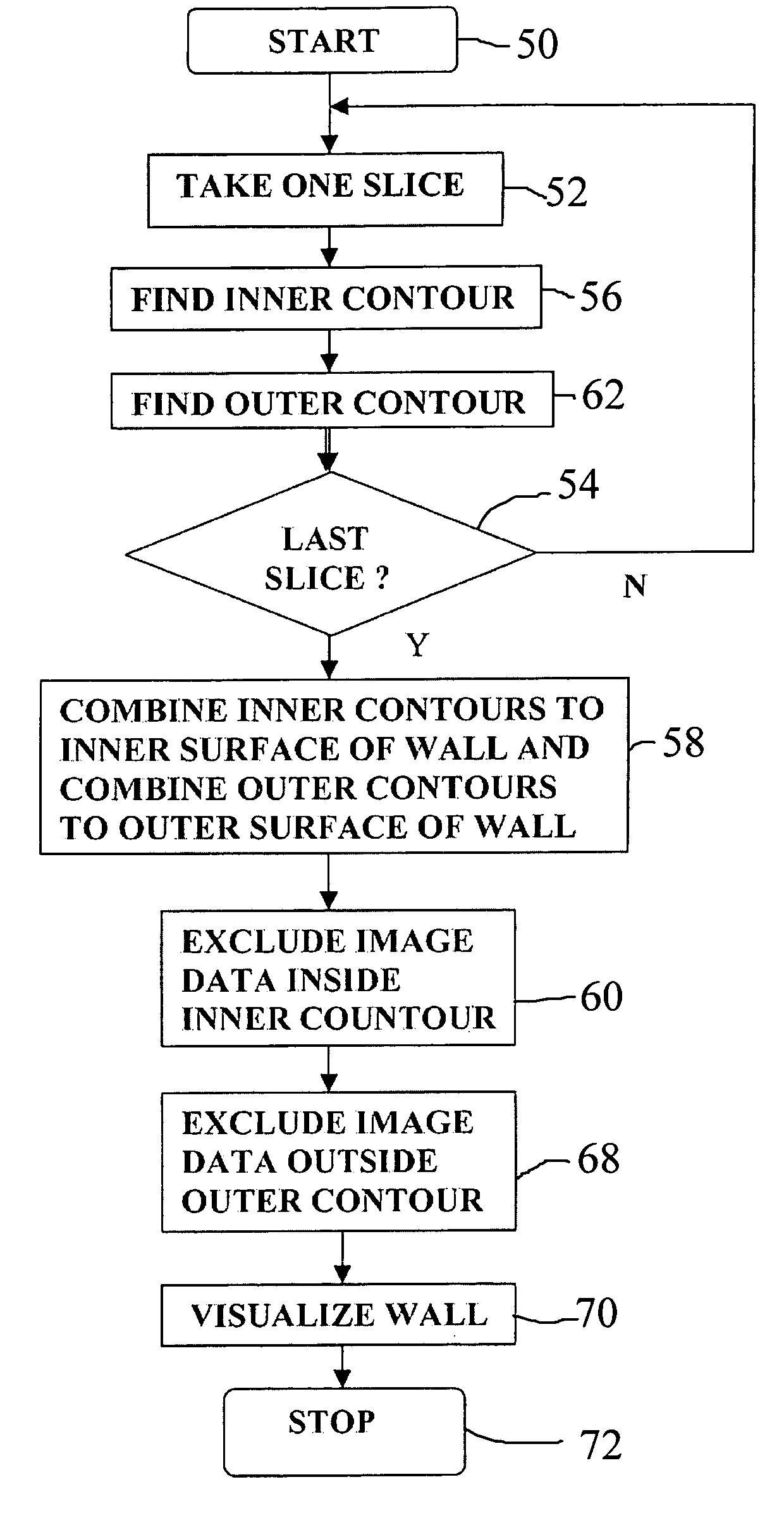
[0016]For the purposes of description, a region between the two contour surfaces will be referred to hereinafter as the wall, such as a heart's wall or shell.

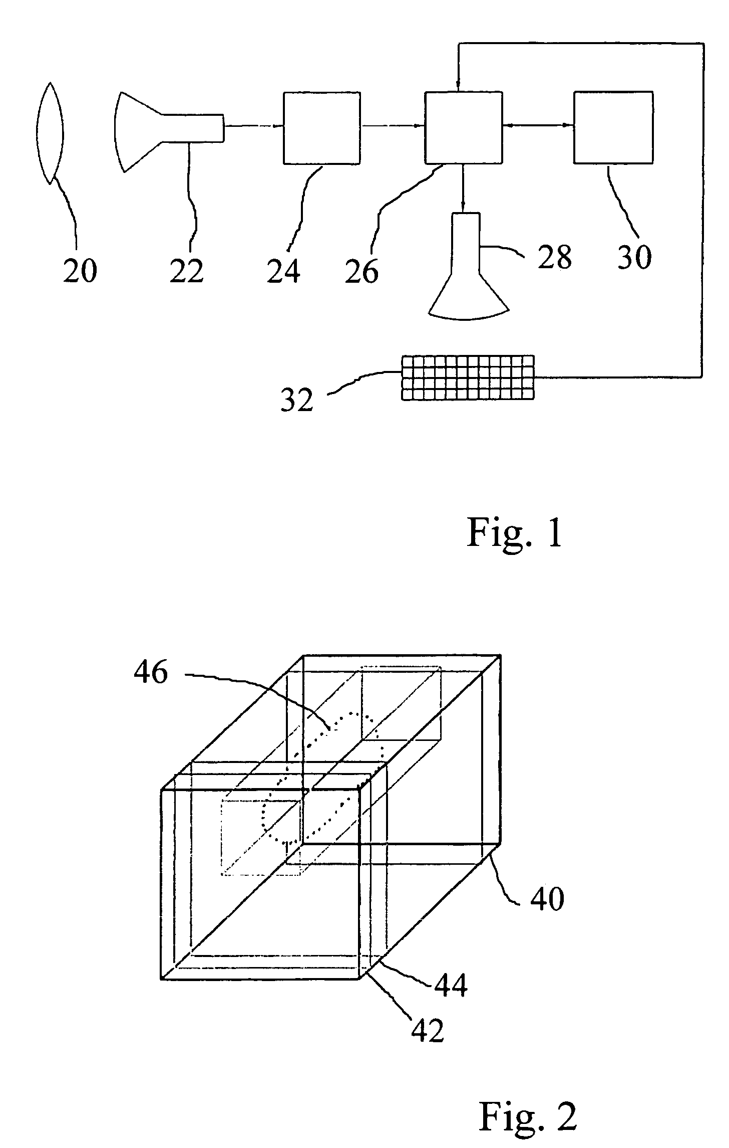
[0017]FIG. 1 illustrates a typical 3D visualizing device. Herein, item 20 is a medical or other biological object, such as a part of a human body. Item 22 is a medical imaging system, such as an X-ray, MR or US device that provides a pattern of relative intensities or other quantitative data, such as represented by a gray-scale. Item 24 symbolizes an image-processing device that may execute various types of image enhancement or other data processing operations. Next, the image is processed in the data processing device 26, thereby resulting in the 3D-image-point (or voxel)-related data set that may subsequently be used for providing a doctor with an appropriate viewing region. These data are transiently stored in storage device 30, and are therein accessed for effecting a display on display screen 28. Through an appropriate use...


PUM


Abstract

A data processing methodology and corresponding apparatus for visualizing the characteristics of a particular object volume in an overall medical / biological environment receives a source image data set pertaining to the overall environment. First and second contour surfaces within the environment are established which collectively define a target object volume. By way of segmenting, all information pertaining to structures outside the target object volume are excluded from the image data. A visual representation of the target object based on nonexcluded information is displayed. In particular, the method establishes i) the second contour surface through combining both voxel intensities and relative positions among voxel subsets, and ii) a target volume by excluding all data outside the outer surface and inside the inner surface (which allows non-uniform spacing between the first and second contour surfaces). The second contour surface is used as a discriminative for the segmenting.

Description

BACKGROUND OF THE INVENTION[0001]1. Field of the Invention[0002]The invention relates to a method for visualizing a particular surface of an object volume in an overall medical / biological environment, through establishing at least a first and a second contour surface within the above environment that collectively define a target object volume and which method allows to selectively exclude information pertaining to structures outside said target object volume.[0003]2. Related Art[0004]Medical / biological MR (magnetic resonance) diagnostics, medical / biological CT (computer tomography) and US (ultrasonic) technologies have all recognized the importance of visualizing only limited target object volumes while excluding volumes out-of-interest. A relevant but non-limiting example is the visualizing of coronary arteries by excluding information outside the heart's wall that could obscure these objects of interest (e.g., in this case both the interior ventricles of the human heart and also s...

Claims


Application Information

Patent Timeline
Patent Type & Authority Patents(United States)
IPC IPC(8): G06T17/00A61B5/055A61B6/03A61B8/00G06T1/00
CPCG06T7/0083G06T15/08G06T2200/04G06T2207/10081G06T2207/10132G06T2207/20144G06T2207/30048G06T2210/41G06T7/12G06T7/194
Inventor VERSTRAELEN, BOUDEWIJN JOSEPH ANGELUSVERSTRAELEN, SEBASTIAAN PAUL
Owner PIE MEDICAL IMAGING
Why Eureka
Try Eureka