
Tomographic reconstruction for x-ray cone-beam scan data
a cone beam and scan data technology, applied in material analysis using wave/particle radiation, instruments, nuclear engineering, etc., can solve the problems of severe cb artifacts, algorithm is not optimal, derived algorithm, not necessarily achieving optimal reconstruction, etc., to suppress cb artifacts and reduce data acquisition
- Summary
- Abstract
- Description
- Claims
- Application Information
AI Technical Summary
Benefits of technology
Problems solved by technology
Method used
Image

Examples
Embodiment Construction
Data Acquisition Geometry
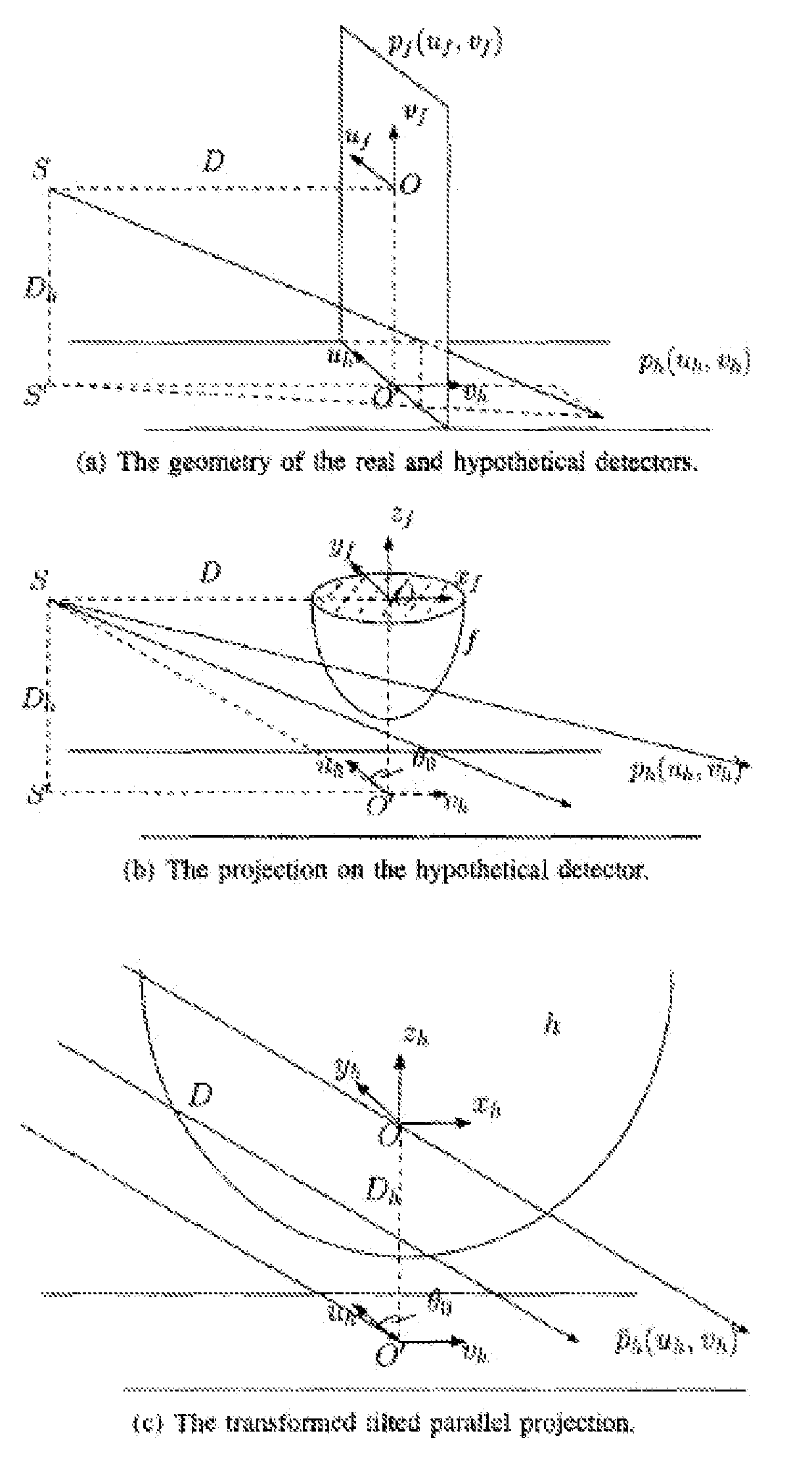
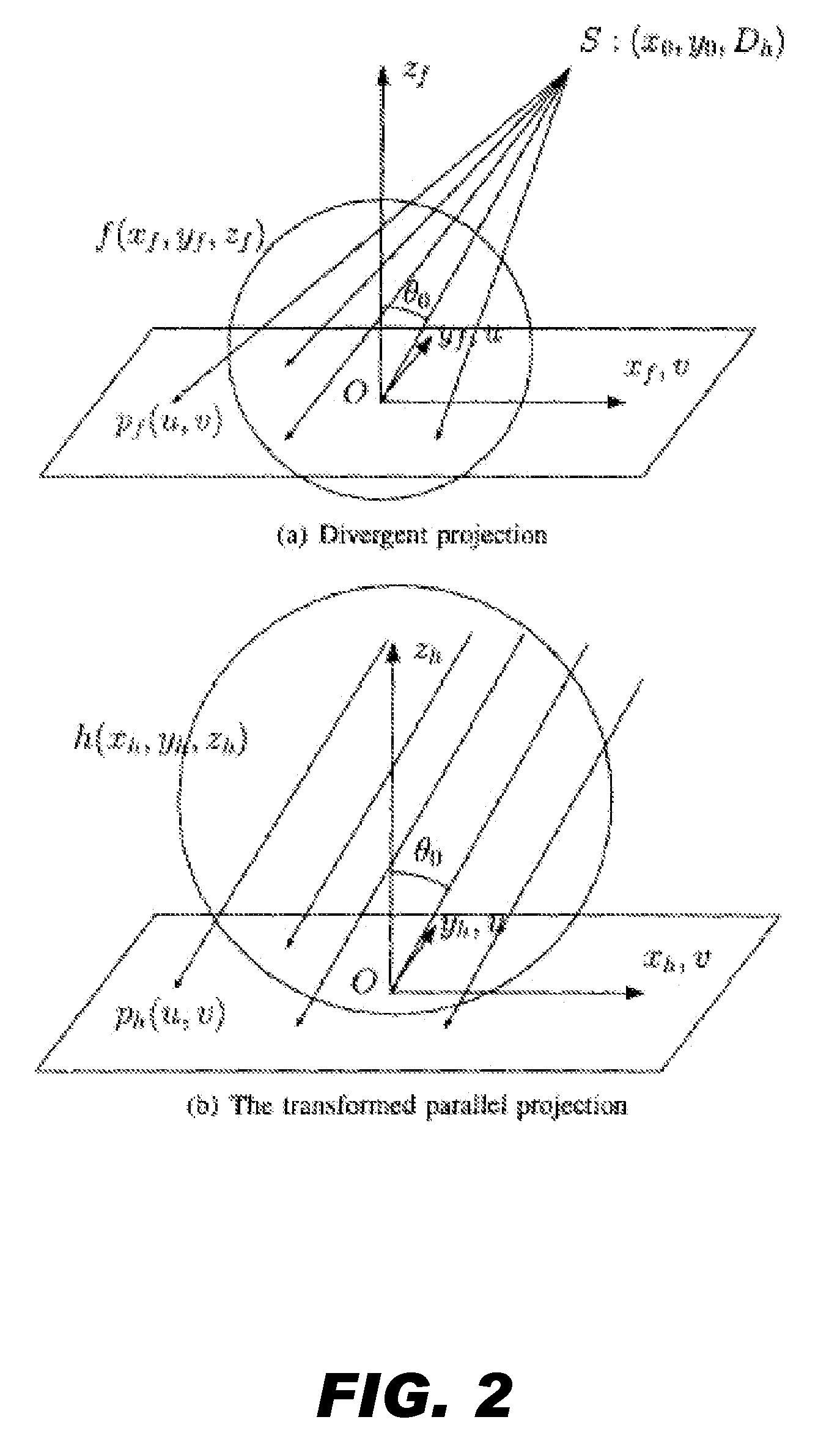
[0017]Consider first a mathematical description of the reconstruction problem, and two theorems that are important in our algorithm derivation. The conversion theorem from divergent projection to parallel projection provides a link between these two imaging geometries, and the FBP-based algorithm on a tilted parallel full scan gives the optimal reconstruction if projection data is not truncated and extrapolation is not allowed. A generalized version of the latter is also derived for later use.
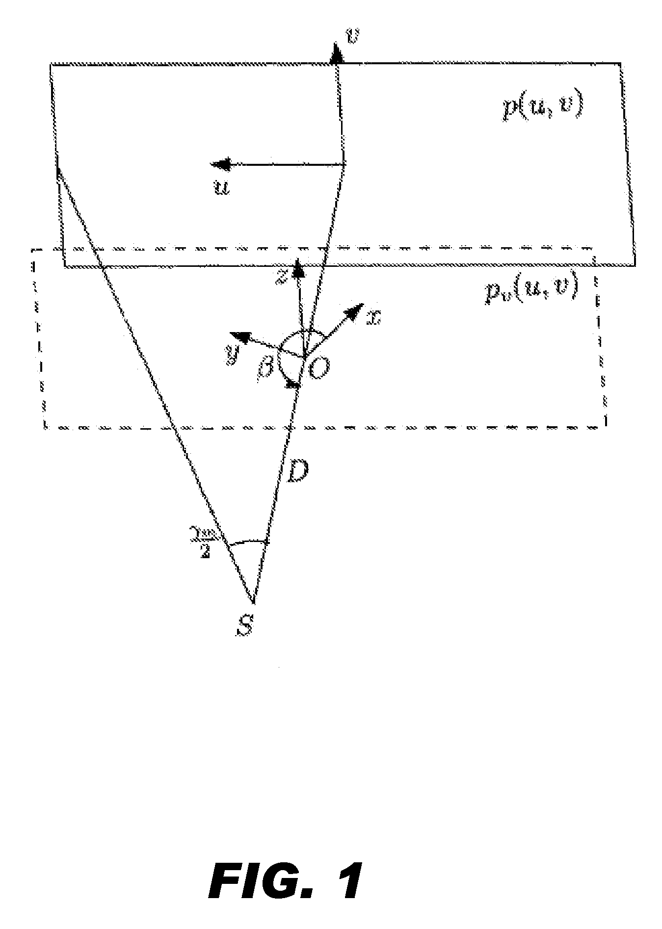
[0018]Herein, we use an equally spaced flat panel detector with a finite size. As shown in FIG. 1, The cone angle γm of this imaging setup is determined by the maximum angular coverage of the cone-beam in the u direction on the detector. During data acquisition, the x-ray source S rotates along the z axis on the x-y plane, with a fixed distance D to the center of rotation O, and the rotation starts at angle βs and stops at βe, with total rotation of not less than π+γm(|...
PUM
| Property | Measurement | Unit |
|---|---|---|
| beam angle | aaaaa | aaaaa |
| attenuation coefficient | aaaaa | aaaaa |
| CT | aaaaa | aaaaa |
Abstract
Description
Claims
Application Information