Sustained delivery of PDGF using self-assembling peptide nanofibers

a peptide nanofiber and self-assembling technology, applied in the direction of peptide sources, peptide/protein ingredients, drug compositions, etc., can solve the problems of simple enmeshed compounds in the peptide matrix, and the tendency to be rapidly lost after implantation, so as to preserve myocardial systolic function, reduce cardiomyocyte death, and high membrane permeability

Inactive Publication Date: 2008-09-30
THE BRIGHAM & WOMEN S HOSPITAL INC
View PDF11 Cites 23 Cited by
  • Summary
  • Abstract
  • Description
  • Claims
  • Application Information

AI Technical Summary

Benefits of technology

[0006]One problem with using membranes formed from self-assembling peptides for drug delivery is that compounds simply enmeshed in the peptide matrix, tend to be rapidly lost after implantation due to the high permeability of membranes. The present invention is based upon the discovery that PDGF, and particularly the B chain of PDGF, binds naturally to self-assembling peptides. This allows biological membranes to be formed that have immobilized PDGF directly attached and which can be implanted in vivo without the PDGF being rapidly washed away. Using a rat model of myocardial injury, membranes with bound PDGF can be locally injected into injured myocardium and are retained at the delivery site for at least 14 days after coronary artery ligation. As a result, cardiomyocyte death is decreased and myocardial systolic function is preserved. Thus, membranes with bound PDGF provide an effective method for preventing heart failure after myocardial infarction.

Problems solved by technology

One problem with using membranes formed from self-assembling peptides for drug delivery is that compounds simply enmeshed in the peptide matrix, tend to be rapidly lost after implantation due to the high permeability of membranes.

Method used


Image

  • Sustained delivery of PDGF using self-assembling peptide nanofibers

Examples


examples

I. Introduction

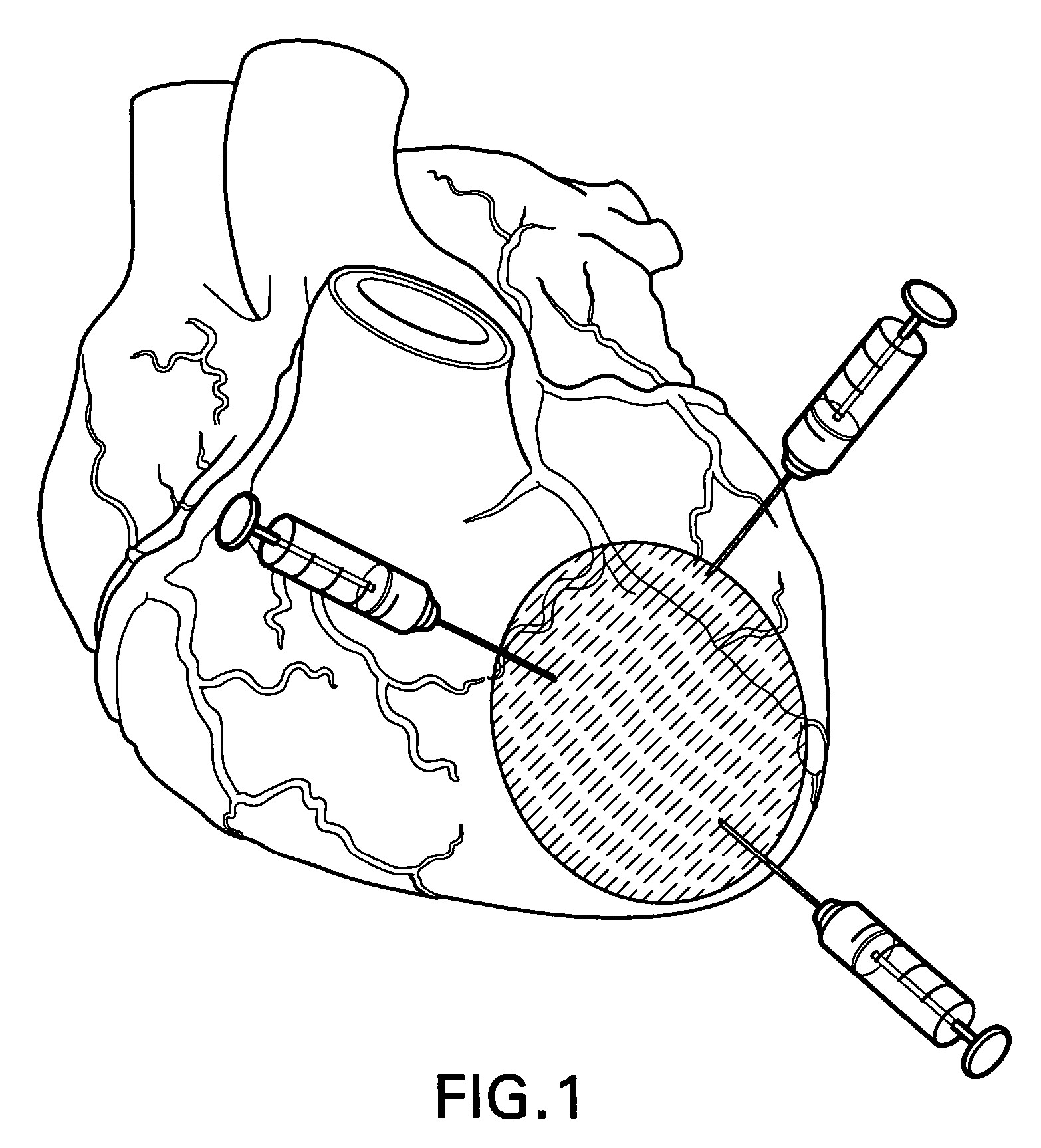
[0035]The present example demonstrates that protection of cardiomyocytes by endothelial cells is through PDGF-BB signaling. PDGF-BB induced cardiomyocyte Akt phosphorylation in a time- and dose-dependant manner and prevented apoptosis via PI3K / Akt signaling. Using injectable self-assembling peptide nanofibers, which bound PDGF-BB in vitro, sustained delivery of PDGF-BB to the myocardium at the injected sites for 14 days was achieved. A blinded and randomized study of 96 rats showed that injecting nanofibers with PDGF-BB, but not nanofibers or PDGF-BB alone, decreased cardiomyocyte death and preserved systolic function after myocardial infarction. A separate blinded and randomized study in 52 rats showed that PDGF-BB delivered with nanofibers decreased infarct size after ischemia-reperfusion. PDGF-BB with nanofibers induced PDGFR-β and Akt phosphorylation in cardiomyocytes in vivo. These data demonstrate that endothelial cells protect cardiomyocytes via PDGF-BB signali...


PUM

PropertyMeasurementUnit
lengthaaaaaaaaaa
concentrationaaaaaaaaaa

Abstract

The present invention is directed to a therapeutic composition in which human PDGF is bound directly to peptides that self assemble into a biologically compatible gel. When implanted in a patient's body, the composition provides for the slow, sustained release of PDGF. The composition will be especially useful in treating patients who have undergone a myocardial infarction.

Description

CROSS REFERENCE TO RELATED APPLICATIONS[0001]The present application claims the benefit of, and priority to, U.S. provisional applications 60 / 640,998, filed Jan. 4, 2005, and 60 / 702,318 filed on Jul. 26, 2005.FIELD OF THE INVENTION[0002]The present invention is directed to a therapeutic membrane composition formed by self-assembling peptides and platelet derived growth factor (PDGF). The membrane / PDGF complex may be implanted or injected into the heart of a patient to provide for the long term release of free PDGF.BACKGROUND OF THE INVENTION[0003]Certain peptides are capable of self assembly when incubated in the presence of a low concentration of monovalent metal cation (U.S. Pat. No. 5,670,483; 6,548,630). Assembly results in the formation of a gel-like membrane that is non-toxic, non-immunogenic and relatively stable to proteases. Once formed, membranes are stable in serum, aqueous solutions and cell culture medium. They can be made under sterile conditions, are capable of suppor...

Claims


Application Information

Patent Timeline
Patent Type & Authority Patents(United States)
IPC IPC(8): A61K38/18
CPCA61K38/10A61K38/1858A61K47/48238C07K7/08C07K14/001C07K14/49A61K2300/00A61K47/62A61P17/02A61P19/08A61P21/00A61P43/00A61P9/10
Inventor LEE, RICHARD T.HSIEH, PATRICK
Owner THE BRIGHAM & WOMEN S HOSPITAL INC
Why Eureka
Try Eureka