

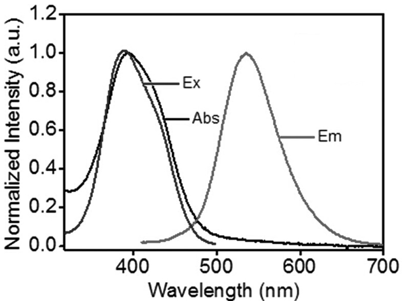
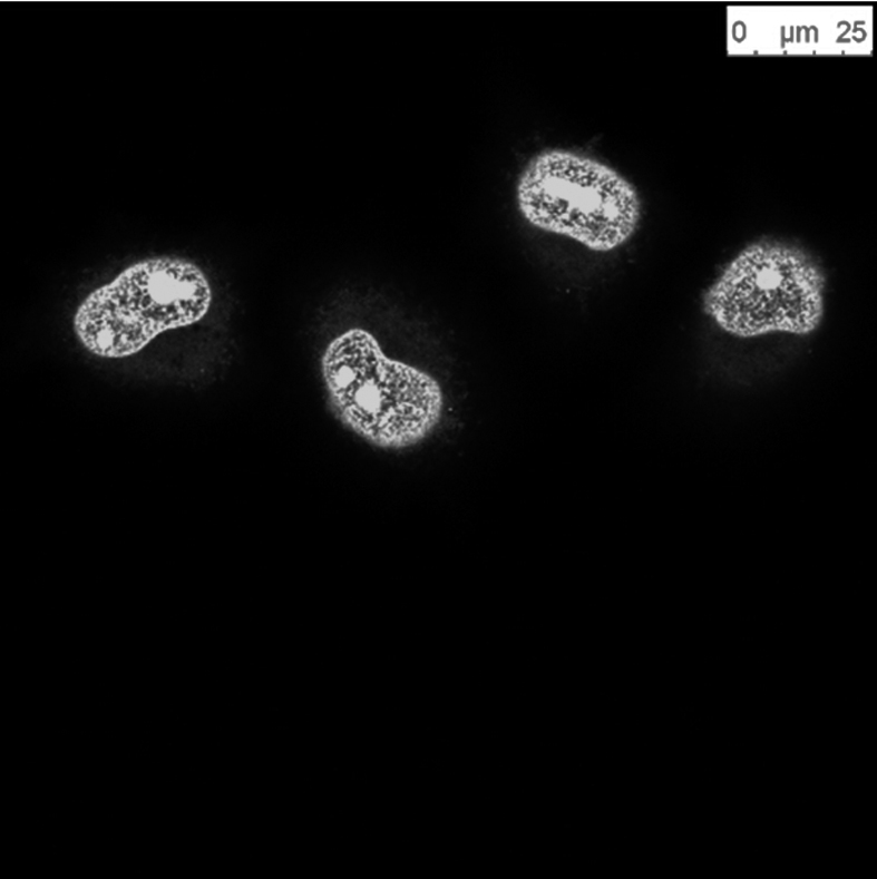
A fluorescent carbon dot for nuclear staining and its application and method in nuclear imaging
A fluorescent carbon dot and fluorescent imaging technology, applied in the field of nanomaterials and biological imaging, can solve the problems of long incubation time and difficult cell nucleus imaging, and achieve the effects of short incubation time, cheap devices and reagents, and low incubation concentration
- Summary
- Abstract
- Description
- Claims
- Application Information
AI Technical Summary
Problems solved by technology
Method used
Image

Examples
Embodiment 1
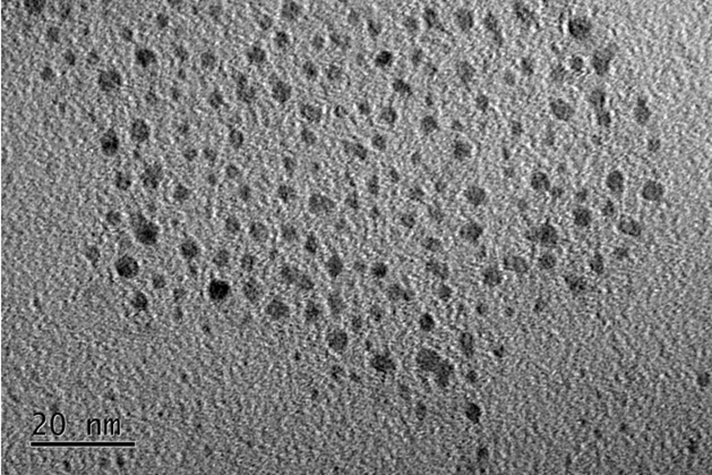
[0029] A kind of fluorescent carbon dots for cell nucleus staining, the carbon dots are prepared by the following steps:
[0030] 1) Dissolve 5 mg of folic acid and 13 mg of m-phenylenediamine in 5000 mg of deionized water, place it in a hydrothermal reactor and heat it to 200 °C for 12 hours; in other embodiments, the mass ratio of folic acid to m-phenylenediamine is 1 : (2-20) can be used, the amount of deionized water can be enough to dissolve the raw materials;
[0031] 2) After the reaction, centrifuge (remove large particle agglomerates) to take the upper layer solution and pass it through silica gel column chromatography to collect the yellow-green fluorescent part, rotate it (remove the organic solvent), transfer it to distilled water, and freeze-dry it to obtain fluorescent carbon dots; The mobile phase used in silica gel column chromatography is a mixture of methanol and ethyl acetate in a volume ratio of 1:1; the specific operation of freeze-drying is: first freeze ...
PUM
| Property | Measurement | Unit |
|---|---|---|
| particle diameter | aaaaa | aaaaa |
| particle size | aaaaa | aaaaa |
Abstract
Description
Claims
Application Information