Patient positioning device of a panoramic dental X-ray apparatus

a positioning device and panoramic technology, applied in dentistry, dental surgery, medical science, etc., can solve the problems of not being able to set standard positions, not allowing height adjustment of the bite block, and being unsuitable for radiographing

Active Publication Date: 2009-08-11
SIRONA DENTAL SYSTEMS
View PDF7 Cites 50 Cited by
  • Summary
  • Abstract
  • Description
  • Claims
  • Application Information

AI Technical Summary

Benefits of technology

[0011]The advantage gained is that the operator does not have to change the positioners for different radiographs but only carries out vertical adjustment. Instead of the three bite blocks used hitherto, i.e. one standard bite element and two bite elements for the midfacial radiographs and accordingly three supporting elements, a single bite block and a single supporting element may be used. This reduces the risk of omitting to change the positioner for body size adjustment.
[0012]Advantageously, the retainer has a socket for fixation on a support of the X-ray device and the longitudinal guide is set axially at a distance from the socket, advantageously in the direction toward the patient. This makes it possible to move the positioner past the carrier during longitudinal displacement.
[0013]Advantageously, detents corresponding to a normal jaw can be provided for the positioner in the longitudinal guide for predefined standard levels. This permits rapid and secure fixation at the correct level.
[0014]In the longitudinal guide for fixation of the positioner, fixing means are advantageously provided in positions lying outside of the detents. By such means it is possible to effect individual adaptations to the anatomical peculiarities of the patient.

Problems solved by technology

As a result of the change, despite all precautionary measures, the situation may arise that a bite block unsuitable for radiographing is used.
This does not allow for any height adjustment of the bite block, however, since only the angle of inclination of the chewing plane is to be considered or the X-ray device has to be adjusted to a pre-assigned value.
However, it sometimes occurs that in the predefined positions when performing transverse tomography (TSA) of the molars, the ramus, i.e. the lower edge of the mandible is not completely imaged.
The disadvantage here is that no standard positions can be set.

Method used


Image

  • Patient positioning device of a panoramic dental X-ray apparatus
  • Patient positioning device of a panoramic dental X-ray apparatus

Examples


Embodiment Construction

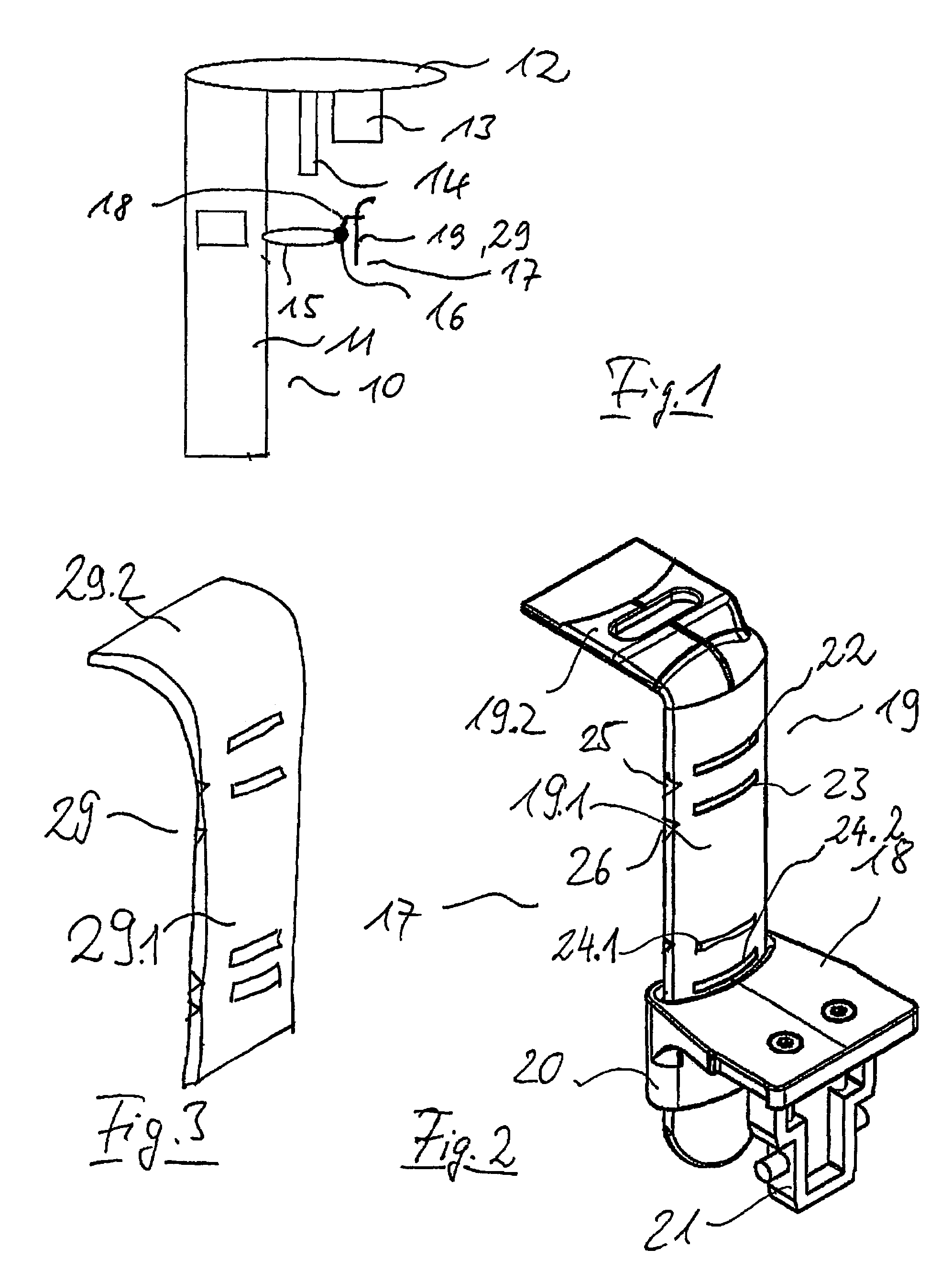
[0021]FIG. 1 is a schematic representation of a panorama X-ray imaging device 10 in which a swiveling unit 12 disposed on a column 11 carries a radiation source 13 and a detector camera 14 located opposite it. On the column 11, in addition, there is a carrier 15 comprising a holder 16 for a positioning device 17 comprising a retainer 18 for a positioner 19 that is vertically adjustable relative to the retainer 18.

[0022]Adaptation of the device 10 to the body size of the patient is accomplished by adjustment of the rotating unit 12, if necessary together with vertical adjustment of the support 15. Adaptation for the type of radiograph is accomplished by adjustment of the positioning device 17.

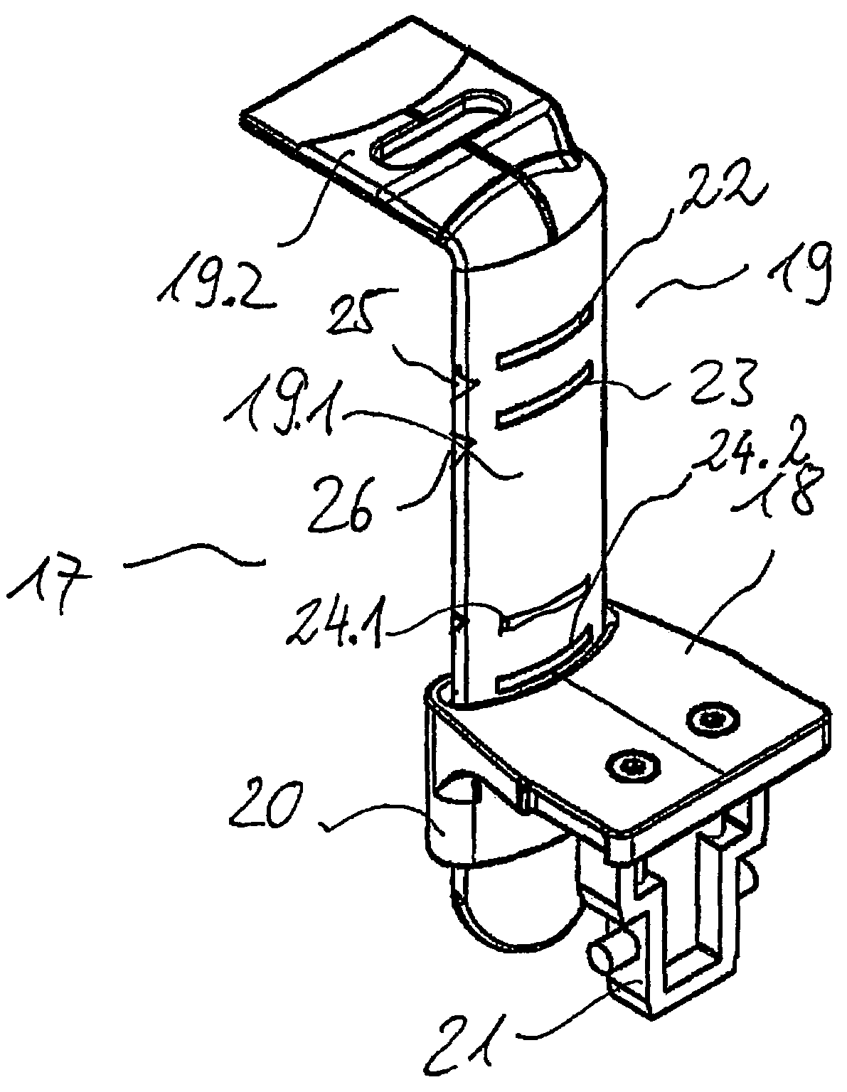
[0023]FIG. 2 is a perspective view of the positioning device 17 according to an exemplary embodiment of the invention comprising a bite block 19. The positioning device 17 has a retainer 18 on which the bite block 19 is guided longitudinally by means of a longitudinal guide 20. The bite block 19...


PUM


Abstract

A positioning device for positioning a patient when being radiographed with a panorama X-ray machine 10 includes a retainer 18 disposed in a fixed direction relative to the X-ray apparatus 10 and a variable positioner connected thereto, for example a bite block 19 or a support, the retainer including a longitudinal guide 20 for guiding the positioner 19 such that the positioner 19 is guided in the retainer 18 for vertical adjustment in a longitudinal direction.

Description

TECHNICAL FIELD[0001]The invention relates to a positioning device for correct positioning of a patient when being radiographed with a panorama X-ray imaging device.PRIOR ART[0002]When a panorama X-ray camera is used, the operator must position the patient prior to take the radiograph and optionally also during the radiographing procedure. This is accomplished with a positioning device comprises a bite block or a positioning support. For different types of radiographs, the positioner must be interchanged in order to permit positioning at different levels. As a result of the change, despite all precautionary measures, the situation may arise that a bite block unsuitable for radiographing is used. Furthermore, for patients having no front teeth, other positioning supports may be necessary, such as a subnasal positioning support that the patient does not bite on but rather against which he rests.[0003]DE 36 09 260 A1 discloses a panorama X-ray device for producing panorama X-ray radiog...

Claims


Application Information

Patent Timeline
Patent Type & Authority Patents(United States)
IPC IPC(8): H05G1/00
CPCA61B6/14A61B6/04
Inventor PRUSS, MALTEDOBERT, MICHAEL
Owner SIRONA DENTAL SYSTEMS
Why Eureka
Try Eureka