Ultrasound target vessel occlusion using microbubbles

a technology of ultrasonic target vessels and microbubbles, which is applied in the field of selective occlusion of blood flow in the vessel, can solve the problems of mechanical damage to the endothelial cells, the damage of the endothelial tissue contacted by the expandable member, and the insufficient size of the fibrin clot to achieve a meaningful occlusion of the blood vessel, etc., and achieve the effect of expanding the original fibrin clo

Inactive Publication Date: 2009-09-22
UNIV OF WASHINGTON
View PDF69 Cites 128 Cited by
  • Summary
  • Abstract
  • Description
  • Claims
  • Application Information

AI Technical Summary

Benefits of technology

[0009]Several different techniques can be used to selectively damage the endothelial cells at a target location where an occlusion is desired. As will be discussed in greater detail below, in a particularly preferred embodiment of the present invention, the endothelial cells are damaged by using a short pulse of HIFU to induce cavitation proximate the target location. Cavitation is an energetic phenomenon, which will result in mechanical damage to the endothelial cells proximate the cavitation sites. This technique has the benefit of substantially eliminating thermal damage to perivascular tissue. It should be understood however, that other techniques can be used to selectively damage endothelial cells proximate the target location.
[0011]While endothelial cells certainly can be selectively damaged using a catheter, using ultrasound facilitates a less invasive technique to be achieved, because ultrasound waves can pass through tissue. Depending on the location of the blood vessel, the ultrasound transducer may be able to be disposed outside of the body entirely. Other blood vessels can be reached by ultrasound transducers disposed within body cavities that can be accessed non-invasively, such as the rectum, vagina, esophagus or stomach.
[0013]A particularly preferred technique for selectively damaging endothelial cells in a blood vessel involves introducing an ultrasound activatable agent into the blood vessel, and using HIFU to activate the ultrasound activatable agent in a very well-defined spatial region corresponding to the target location. The ultrasound activatable agent disposed in portions of the blood vessel not corresponding to the focal region of the HIFU beam will not be activated, thus the cavitational damage will be limited to the portion of the blood vessel corresponding to the focal region of the HIFU beam. A useful ultrasound activatable agent are microbubbles, and ultrasound contrast agents represent particularly preferred types of the microbubbles. Because the ultrasound activatable agent will only be activated in the focal region of the HIFU beam, the ultrasound activatable agent can be introduced into the blood vessels systemically, or directly into the blood vessel in question (for example, by direct injection or using a delivery catheter). Significantly, only a very short burst of HIFU is required to activate such ultrasound activatable agents, which means that the duty cycle can be made to be of sufficiently short duration that a negligible amount of acoustic energy is absorbed by tissue in the focal region of the HIFU beam. Thus, even if some perivascular tissue lies within the focal region of the HIFU beam, no (or minimal) thermal damage will occur to the perivascular tissue, because the HIFU beam will not be energized long enough for the perivascular tissue to absorb enough acoustic energy to damage the perivascular tissue.

Problems solved by technology

Generally, the resulting fibrin clot will not be sufficiently large to achieve a meaningful occlusion of the blood vessel.
Cavitation is an energetic phenomenon, which will result in mechanical damage to the endothelial cells proximate the cavitation sites.
An expandable member portion of the catheter is then inflated such that the endothelial tissue contacted by the expandable member is damaged.

Method used


Image

  • Ultrasound target vessel occlusion using microbubbles
  • Ultrasound target vessel occlusion using microbubbles
  • Ultrasound target vessel occlusion using microbubbles

Examples


Embodiment Construction

Overview of the Present Invention

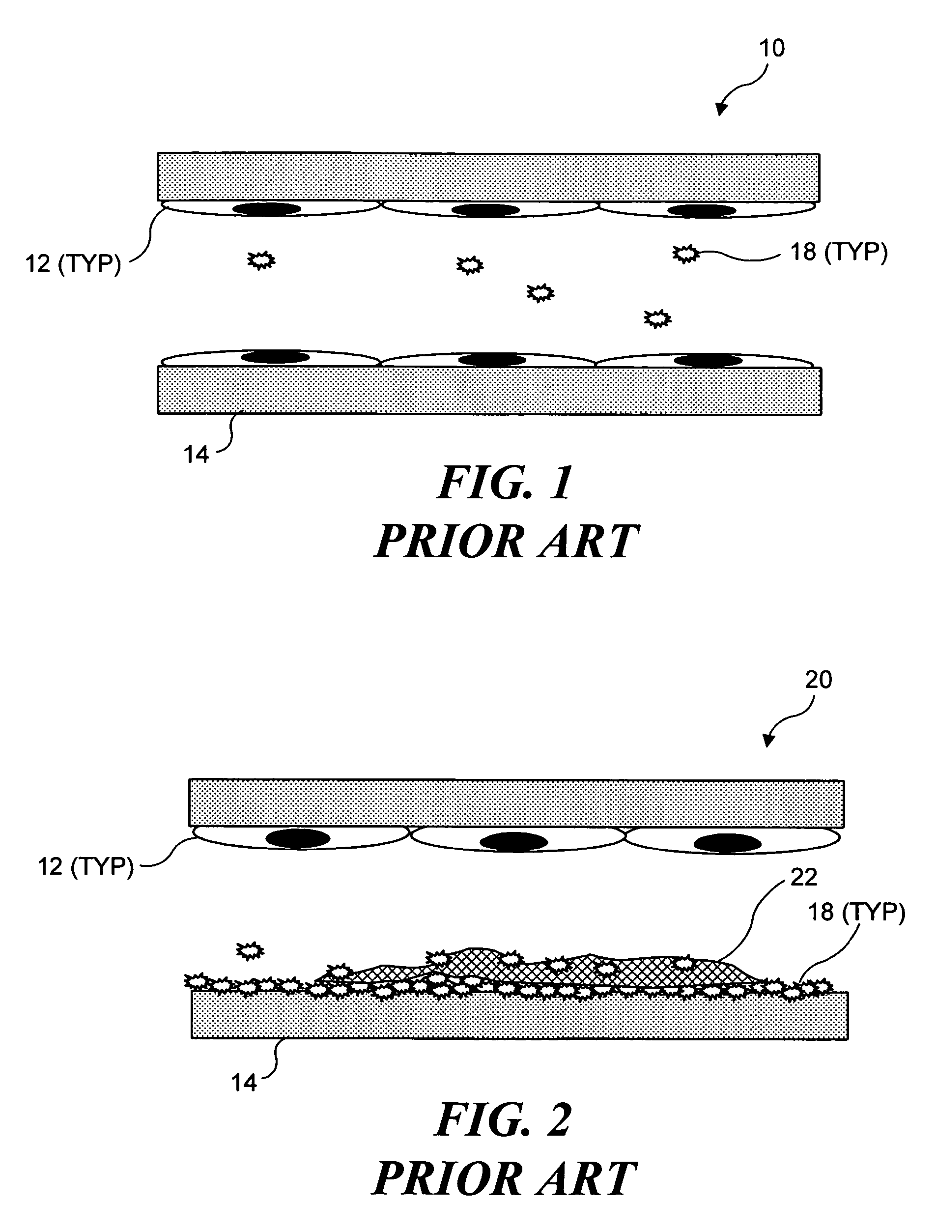
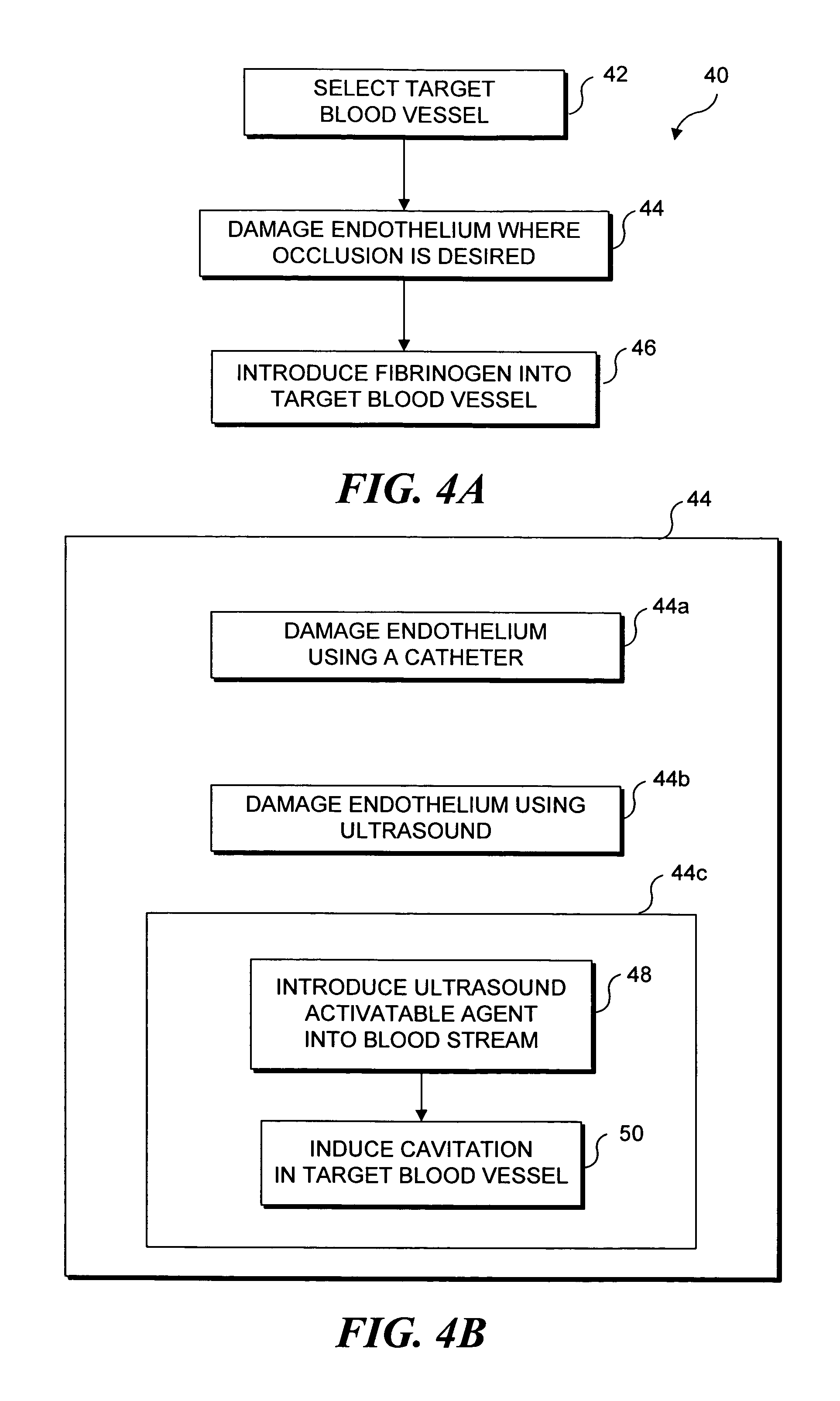
[0033]As noted above, the present invention can be used to selectively occlude a blood vessel by selectively damaging endothelial cells in a portion of a blood vessel where an occlusion is desired. The damage to the endothelial cells exposes underlying tissue, which releases enzymes causing naturally occurring fibrinogen to polymerize, thereby depositing a fibrin clot along the vessel wall. Additional fibrinogen is introduced into the blood vessel. The additional fibrinogen is similarly converted to fibrin, enlarging the clot. An advantage of the present invention is that because the thrombus is formed on the underlying tissue where damage was caused to the endothelial tissue, the clot that is produced cannot readily break loose from that site and cause undesirable problems in other parts of the body, e.g., by occluding a blood vessel in a patient's brain. While a number of different techniques can be used to selectively damage the endothelial cells,...


PUM

PropertyMeasurementUnit
temperaturesaaaaaaaaaa
lengthaaaaaaaaaa
diameteraaaaaaaaaa

Abstract

Selective occlusion of a blood vessel is achieved by selectively damaging endothelial cells at a target location in the blood vessel, resulting in the formation of a fibrin clot proximate to the damaged endothelial cells. Additional fibrinogen can then be introduced into the blood vessel if occlusion is not achieved, as the fibrinogen is converted to fibrin by enzymes released by the exposed thrombogenic tissue and activated platelets. Endothelial cells are selectively damaged using thermal effects induced by ultrasound, by mechanical effects induced by ultrasound, or by mechanical effects produced by a tool introduced into the blood vessel (such as a catheter-based tool). A particularly preferred technique for selectively damaging endothelial cells involves introducing an ultrasound activatable agent into the blood vessel, and causing cavitation in that agent using pulses of high-intensity focused ultrasound having a duration insufficient to induce thermal damage in adjacent perivascular tissue.

Description

[0001]This invention was made with U.S. Government support under grants 1F32DK65413-01 and 8R01-EB00350-02 awarded by the National Institutes of Health, and U.S. Army Contract No. DAMD17-02-2-0014, awarded by the Department of Defense. The U.S. Government has certain rights in the invention.FIELD OF THE INVENTION[0002]The present invention generally relates to selectively occluding blood flow in a vessel, and more specifically, to a method of selectively occluding blood flow by damaging endothelial cells at a portion of the blood vessel in which an occlusion is desired, and then introducing fibrinogen into the blood vessel.BACKGROUND OF THE INVENTION[0003]While it is widely recognized that reduction of blood flow in arties is a major factor in heart disease, there are many medical conditions in which restricting or occluding blood flow through a blood vessel is desirable. For example, sclerotherapy is the injection of a sclerosing agent (such as morrhuate sodium) into a blood vessel...

Claims


Application Information

Patent Timeline
Patent Type & Authority Patents(United States)
IPC IPC(8): A61B17/26A61B8/00A61B8/12A61K38/00
CPCA61N7/02A61K38/363A61B8/0833A61B2017/22008A61M2025/109
Inventor HWANG, JOO HABRAYMAN, ANDREW
Owner UNIV OF WASHINGTON
Why Eureka
Try Eureka