Devices and methods for interstitial injection of biologic agents into tissue

a technology of biologic agents and devices, applied in the field of medical devices and methods, can solve the problems of unattractive scars, increased complications, and severe trauma to thoracic muscles and ribs

Active Publication Date: 2009-12-08
MEDTRONIC INC
View PDF761 Cites 71 Cited by
  • Summary
  • Abstract
  • Description
  • Claims
  • Application Information

AI Technical Summary

Benefits of technology

[0037]In still another device, the first body has a plurality of hollow needles attached thereto and a first plurality of magnets secured thereto. The second body has a plurality of needles slidably received therethrough. A third body can be slidably disposed on the second body and have a second plurality of magnets thereon, where the first and second plurality of magnets are disposed and have polarities oriented such that the slidable third body has a first position in which the first and third body are magnetically attracted to each other and a second position in which the first and third body are magnetically repulsed from each other. In this way, sliding the third member can pull the first body toward the second body and also push the first body away from the second body, depending on the degree of sliding. In one device, the third body has longitudinally adjacent magnetic pairs having opposite polarities, such that sliding the third member into the first position brings magnets having opposite facing polarities opposite each other, and sliding the third member into a second position brings magnets having the same facing polarities opposite each other.
[0040]In some devices, the plurality of needles attached to the first body may have a substantially different length as among the needles, to form a phased depth array of needles. The phased array of needles can distribute the initially higher force required to puncture the outer tissue temporally over the needle insertion process. In addition, the phased array of needles can allow the delivery one or more medical agents or substances at different depths within the tissue simultaneously.

Problems solved by technology

As a result, these operations typically require large numbers of sutures or staples to close the incision and 5 to 10 wire hooks to keep the severed sternum together.
Such surgery often carries additional complications such as instability of the sternum, post-operative bleeding, and mediastinal infection.
The thoracic muscle and ribs are also severely traumatized, and the healing process results in an unattractive scar.
Problems may develop during CPB due to the reaction blood has to non-endothelially lined surfaces, i.e. surfaces unlike those of a blood vessel.
This may, in turn, increase the risk of hemorrhage.
Therefore, much effort has been expended to develop medical devices and techniques to access the pericardial space employing such minimally invasive percutaneous procedures.
One difficulty has been that normally the pericardial space is so small or thin that it is difficult to penetrate the pericardium using miniaturized instruments capable of being introduced through a port to the site without also puncturing the underling epicardium and thereby, damaging the myocardium or a coronary vessel.
Proliferative adhesions occur between the pericardium and the epicardium in diseased hearts and hamper access to the pericardial space employing such minimally invasive percutaneous procedures.
It is difficult to introduce and use the forceps through the narrow lumen of a port or sleeve, particularly if the pericardial fluid is under pressure that makes the pericardium taut like an inflated balloon.
Although theoretically plausible, the ability to reliably maintain a vacuum seal against the pericardium when such treatment devices are advanced can be problematic.
Heart disease, including myocardial infarction, is a leading cause of death and impaired activity in human beings, particularly in the western world, most particularly among males.
Heart disease can in turn degrade other physiological systems.
A totally blocked, or substantially blocked coronary artery can cause immediate, intermediate term, and long-term problems.
In the immediate term, myocardial cells can be starved of oxygen resulting in cell death.
In the long term, the myocardial cell death, which creates weakened, non-contracting infarct regions of the heart, can lead to heart failure.
One reason is the substantial expense and small amount of the medical agents available, for example, agents used for gene therapy.
Yet another reason is the undesirability or impossibility of systemically delivering agents to the heart tissue requiring treatment.
Open chest procedures are inherently traumatic procedures with associated risks.
The risks are often justified when the alternatives are a substantially high probability of death.
In many cases, however, an open chest procedure is not believed justifiable only for the injection of medical agents into the myocardium.
This approach may not allow all areas of the heart to be easily reached however.
The size and type of instruments that can be advanced, for example, from a femoral artery approach, are also limited.
Current intravascular delivery devices are less than optimal, being limited in the cardiac regions they can reach and the amount and types of materials they can deliver.
An open chest procedure may not be justifiable, however, only for the injection of such cells.
In particular, patients having suffered a recent heart attack may be very poor candidates for such a procedure.

Method used


Image

  • Devices and methods for interstitial injection of biologic agents into tissue
  • Devices and methods for interstitial injection of biologic agents into tissue
  • Devices and methods for interstitial injection of biologic agents into tissue

Examples


Embodiment Construction

[0116]The following detailed description should be read with reference to the drawings, in which like elements in different drawings are numbered identically. The drawings, which are not necessarily to scale, depict selected embodiments and are not intended to limit the scope of the invention. Several forms of invention have been shown and described, and other forms will now be apparent to those skilled in art. It will be understood that embodiments shown in drawings and described below are merely for illustrative purposes, and are not intended to limit the scope of the invention as defined in the claims that follow.

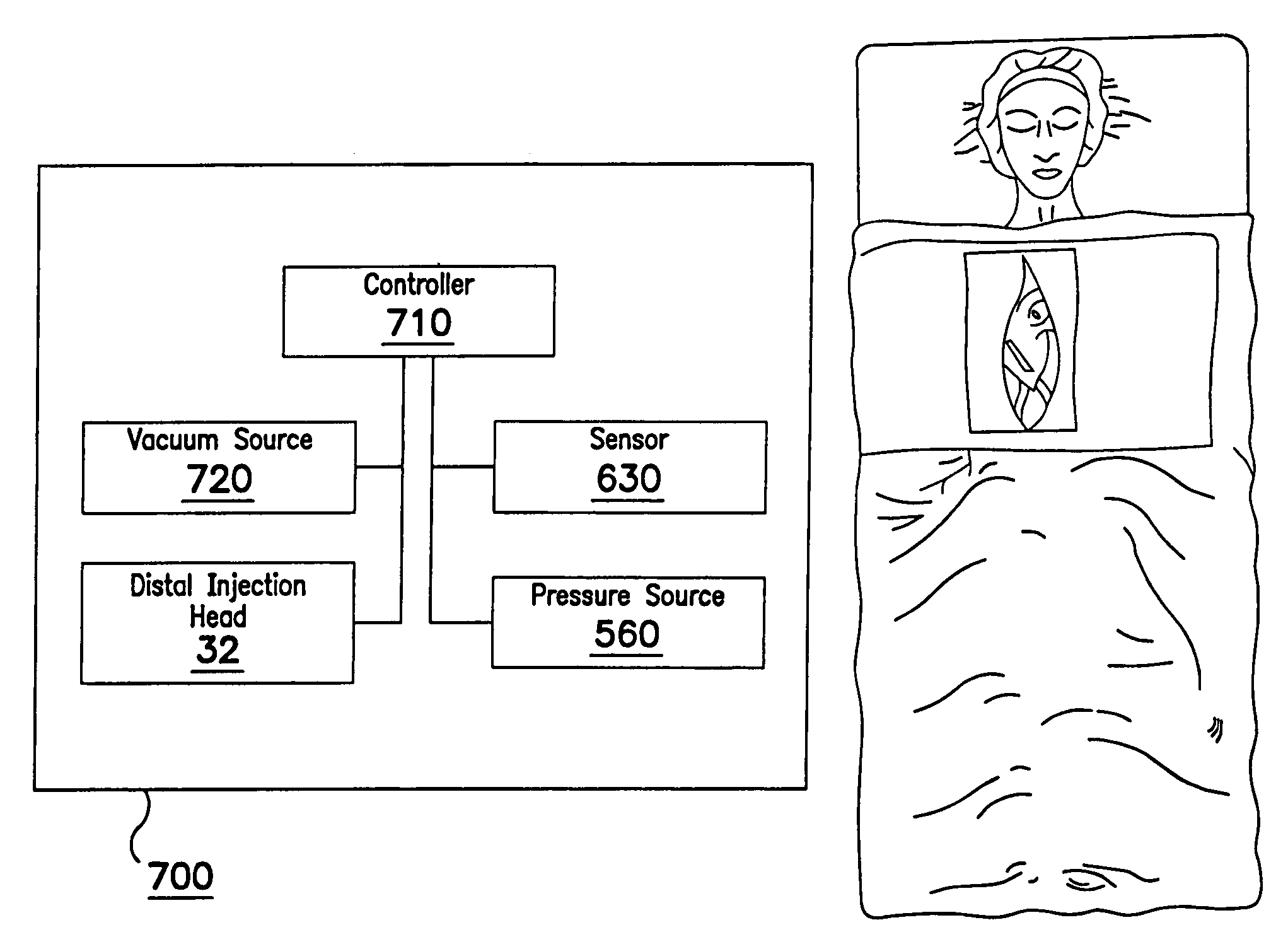
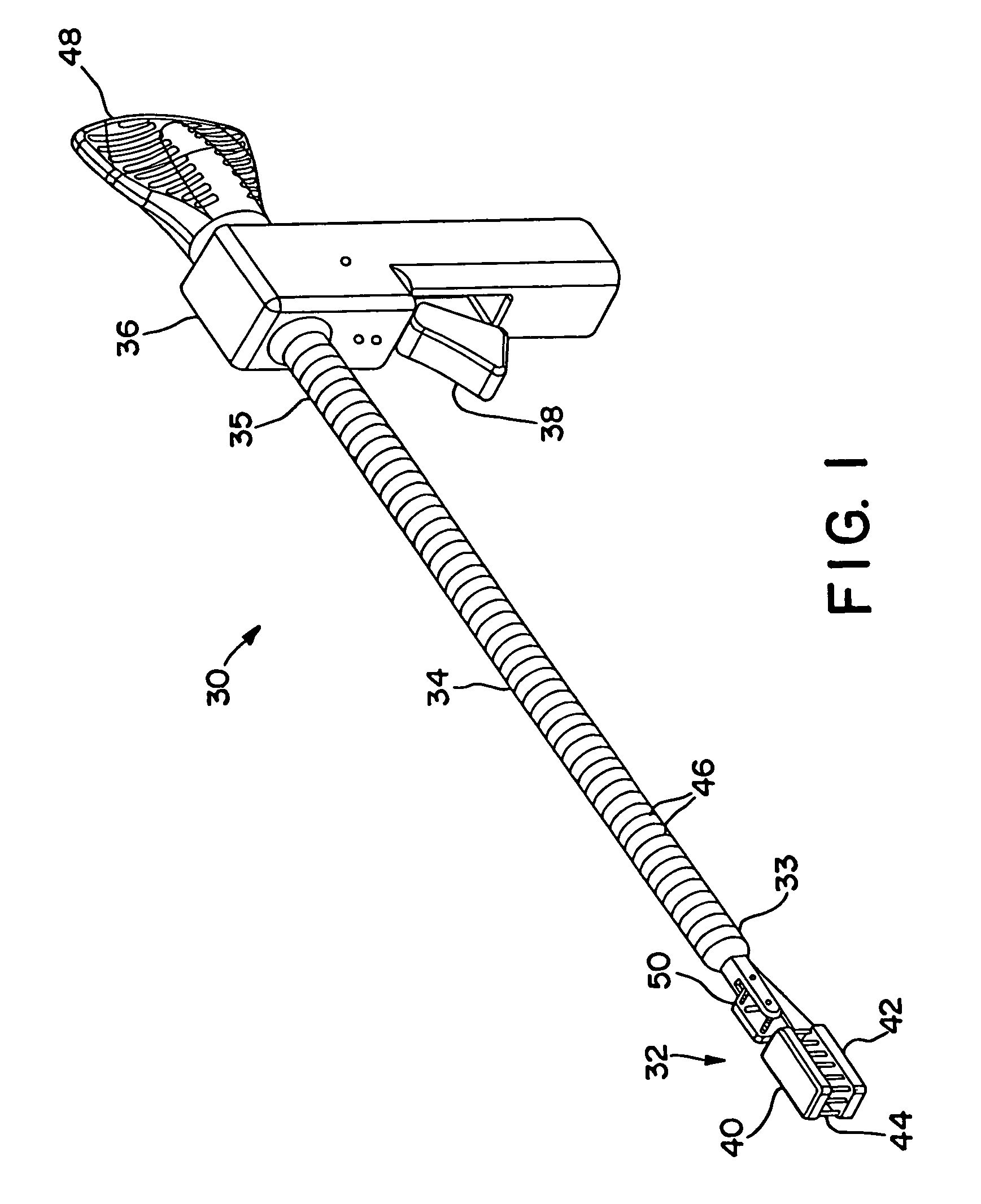
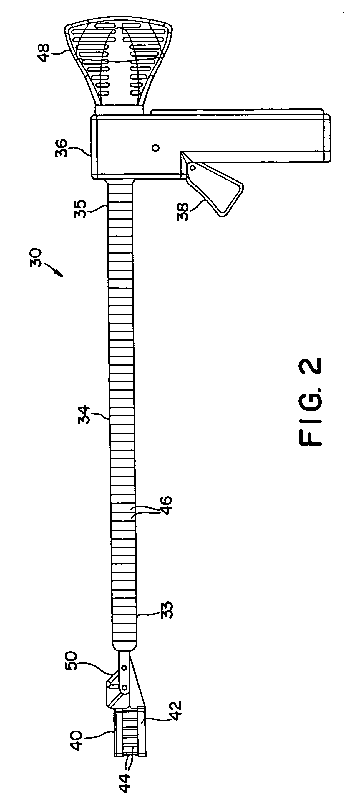
[0117]FIGS. 1 and 2 illustrate an interstitial injection device 30 having a distal injection head 32, an elongate shaft 34, and a proximal handle 36. Elongate shaft 34 includes generally a distal region 33 and a proximal region 35. Proximal handle 36 includes a trigger mechanism 38 for initiating the needle insertion and / or fluid injection. Distal injection head 32 may i...


PUM


Abstract

Apparatus and methods for injecting biological agents into tissue. Devices are provided having elongate shafts and distal injection heads for driving needles into tissue and injecting medical agents into the tissue through the needles. A longitudinal force directed along the shaft can be translated to a needle driving force. Some devices provide controllably variable needle penetration depth. Devices include mechanical needle drivers utilizing four link pantographs, rack and pinions, and drive yokes for driving a first needle bearing body toward a second tissue contacting body. Other devices include inflatable members for driving and retracting needles. Still other devices include magnets for biasing the needles in extended and / or retracted positions. The invention includes minimally invasive methods for epicardially injecting cardiocyte precursor cells into infarct myocardial tissue.

Description

CROSS-REFERENCE TO RELATED APPLICATIONS[0001]This application is a continuation-in-part of U.S. patent application Ser. No. 10 / 341,743, filed Jan. 14, 2003, the disclosure of which is incorporated herein by reference.[0002]This application is also a continuation-in-part of U.S. patent application Ser. No. 10 / 622,147, filed Jul. 17, 2003, now U.S. Pat. No. 6,918,908, which is a continuation-in-part of U.S. patent application Ser. No. 10 / 342,932, filed Jan. 15, 2003, now U.S. Pat. No. 6,837,848, which is related to commonly assigned U.S. patent application Ser. No. 10 / 283,794, filed Oct. 30, 2002, for METHODS AND APPARATUS FOR ACCESSING AND STABILIZING AN AREA OF THE HEART in the names of Gary W. Guenst et al., now U.S. Pat. No. 7,146,225; U.S. patent application Ser. No. 10 / 342,960 filed Jan. 15, 2003, for METHODS AND TOOLS FOR ACCESSING AN ANATOMIC SPACE in the name of Gary W. Guenst, now U.S. Pat. No. 6,786,898; and U.S. patent application Ser. No. 10 / 284,771 filed Oct. 31, 2002, f...

Claims


Application Information

Patent Timeline
Patent Type & Authority Patents(United States)
IPC IPC(8): A61M31/00A61B17/32A61B17/00A61B17/20A61B17/28A61B17/30A61B17/34A61M5/00
CPCA61B17/3478A61B17/00234A61B17/205A61B2017/00243A61B2017/00247A61M25/0084A61B2017/2936A61B2017/2943A61B2017/306A61B2018/00392A61B2019/304A61B2017/2926A61B2090/034
Inventor BONNER, MATTHEW D.ROTHSTEIN, PAUL T.HINIDUMA-LOKUGE, PRASANGA D.KEOGH, JAMES R.USHER, RAYMOND W.JAHNS, SCOTT ERICCHEN, VICTOR T.
Owner MEDTRONIC INC
Why Eureka
Try Eureka