Breast tomosynthesis with display of highlighted suspected calcifications

a tomosynthesis and suspected calcification technology, applied in the field of xray tomosynthesis, can solve the problems of inability to fully appreciate or utilize the information in the images, the risk of some calcifications being noisy, and the examination time becoming so long as to be impractical, etc., to facilitate visualization, enhance all microcalcifications, and be useful to health professionals

Active Publication Date: 2009-12-08
HOLOGIC INC
View PDF22 Cites 142 Cited by
  • Summary
  • Abstract
  • Description
  • Claims
  • Application Information

AI Technical Summary

Problems solved by technology

Unless suitable display techniques are developed, information that is in the images may not be fully appreciated or utilized and / or the examination time may become so long as to be impractical, particularly for screening asymptomatic patients but also possibly for diagnostic or other purposes.
Identifying all of the very smallest calcification-like objects runs the risk that some of them might represent noise and not true calcifications or reasonably suspected calcifications.

Method used


Image

  • Breast tomosynthesis with display of highlighted suspected calcifications
  • Breast tomosynthesis with display of highlighted suspected calcifications
  • Breast tomosynthesis with display of highlighted suspected calcifications

Examples


Embodiment Construction

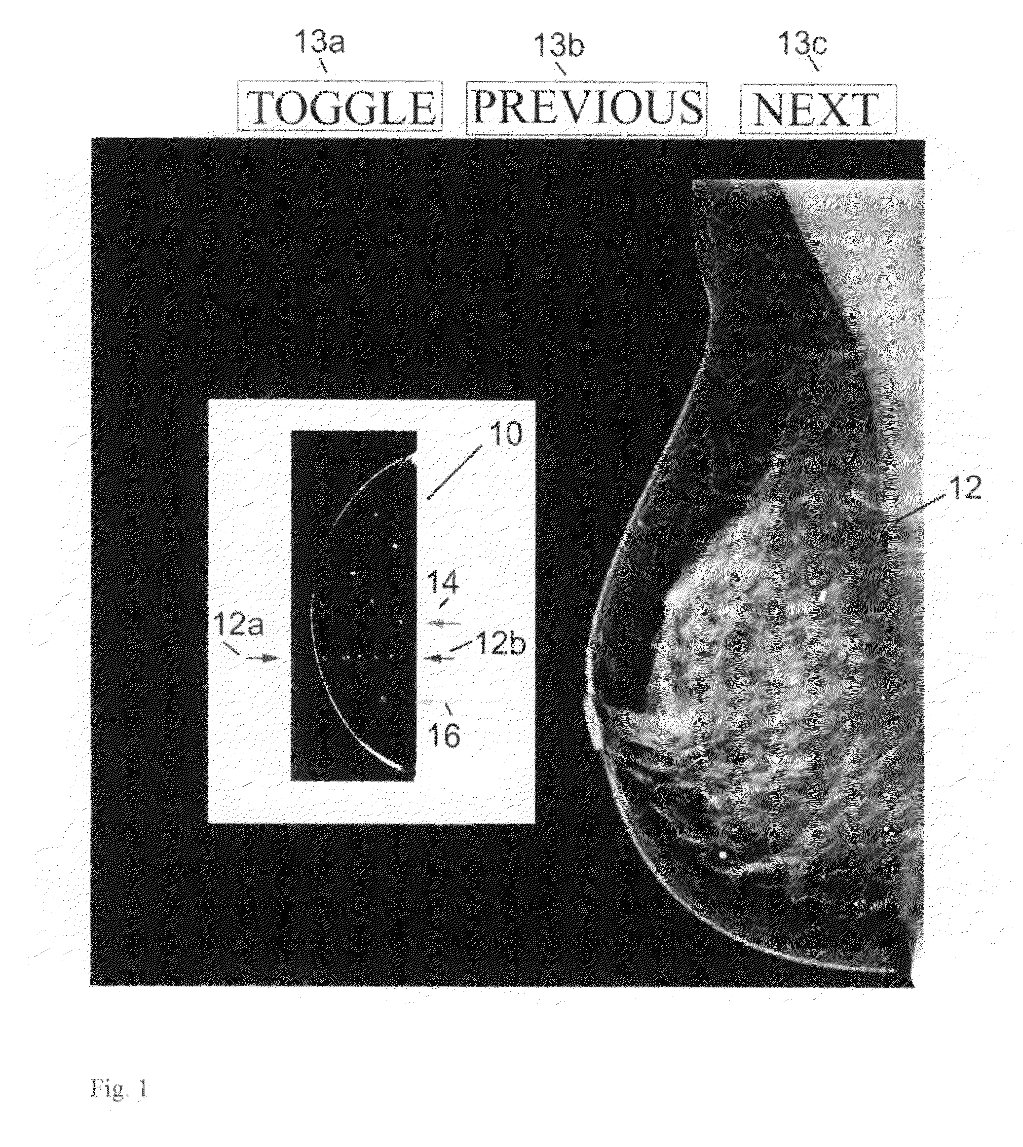
[0019]Referring to FIG. 1, the image on the left is a scout view 10 that generally conforms to an outline of a patient's breast and contains bright dots indicative of calcifications identified through a process described below. Two facing arrows 12a and 12b at the side of scout image 10 point to a level in the scout image that corresponds to a reconstructed tomosynthesis slice image Tr 12 seen at the right side of FIG. 1. The views of images 10 and 12 are mutually orthogonal. Tr image 12 in this example has highlighted calcifications (seen as white dots) that are more numerous than those seen in scout image 10, for reasons that will become apparent from the disclosure below. Next to scout image 10 are an arrow 14 that points to the next level up in image 10 that contains calcifications and an arrow 16 that points to the next level below in image 10 that contains calcifications. By clicking on arrow 14 or arrow 16 the user can call for display at 12 the corresponding Tr image. Tr ima...


PUM


Abstract

Systems and methods that facilitate the presentation and assessment of selected features in projection and / or reconstructed breast images, such as calcifications that meet selected criteria of size, shape, presence in selected slice images, distribution of pixels that could be indicative of calcification relative to other pixels or of other image features of clinical interest.

Description

REFERENCE TO RELATED APPLICATIONS[0001]This application is related to earlier-filed application Ser. Nos. 11 / 604,069, 11 / 271,050, 11 / 059,282, 10 / 723,486, and 10 / 305,480 filed, respectively, on Nov. 24, 2006, Nov. 10, 2005, Feb. 16, 2005, Nov. 26, 2003, and Nov. 27, 2002, and is related to PCT applications PCT / US2005 / 41941 and PCT2005 / 42613 filed, respectively, on Nov. 15 and 23, 2005. This patent specification hereby incorporates by reference the contents of each of the earlier-filed patent applications.FIELD[0002]This patent specification pertains to x-ray tomosynthesis, and more specifically to techniques and equipment for acquiring, processing, storing and displaying tomosynthesis images, including tomosynthesis projection images and tomosynthesis reconstructed images. An important aspect of this patent specification pertains to identifying and highlighting suspected calcifications in displays of tomosynthesis images.BACKGROUND AND SUMMARY OF THE DISCLOSURE[0003]X-ray mammography...

Claims


Application Information

Patent Timeline
Patent Type & Authority Patents(United States)
IPC IPC(8): G06K9/00
CPCG06K9/4638G06T7/0012G06K2209/053G06T2207/30068G06T2207/10081A61B6/502A61B6/025A61B6/12A61B6/461A61B6/5211A61B6/5223A61B6/5252G16H50/20G06V10/457G06V2201/032
Inventor RUTH, CHRISSTEIN, JAYNIKLASON, LORENSMITH
Owner HOLOGIC INC
Why Eureka
Try Eureka