System and method for ultrasound therapy using grating lobes

a technology of grating lobes and ultrasound therapy, applied in the field of ultrasound, can solve the problems of no other known modality that offers noninvasive, no treatment of extensive targets, and consequently no treatment of extensive targets, and achieve the effect of achieving more efficiently

Active Publication Date: 2010-10-05
CILAG GMBH INT
View PDF366 Cites 66 Cited by
  • Summary
  • Abstract
  • Description
  • Claims
  • Application Information

AI Technical Summary

Benefits of technology

[0011]One embodiment of the present invention overcomes the aforementioned limitations of ultrasound transducers by altering the phasing of transducer elements on a transducer array, such as a linear ultrasound array, so that the positions of grating lobes emitted by the array can be changed. A volume of target tissue can be ablated using a sequence of several different insonifications. Each of these insonifications creates multiple grating lobe foci to cover a different subset of the volume. In this manner, ablation of a large target tissue can be accomplished more efficiently than is possible using a single focus without grating lobes.
[0013]Another embodiment of the present invention is a system for medical treatment of tissue using ultrasound. The system comprises a probe having an array of transducer elements; an ultrasound waveform generator adapted to generate at least one electrical ultrasound signal; and a plurality of phase controls coupled to the ultrasound waveform generator, each phase control adapted to generate from the at least one electrical ultrasound signal a drive signal that is coupled to an associated transducer element. The phase controls comprise a focusing phase shift portion adapted to produce a first phase shift in the drive signal, and a grating lobe phase shift portion adapted to produce a second phase shift in the drive signal, wherein each drive signal has a composite phase shift that is the mathematical sum of the first and second phase shifts. The drive signals are effective to cause the array to emit acoustic ultrasound signals having a focal point controlled by the first phase shift and grating lobe foci controlled by the second phase shift.

Problems solved by technology

The absorption of the ultrasonic energy at the focus induces a sudden temperature rise of tissue, which causes ablation of the target volume of cells in the focal region.
Currently there is no other known modality that offers noninvasive, deep, localized focusing of non-ionizing radiation for therapeutic purposes.
Treatment of extensive targets is consequently a problem.
A particular problem is the use of small ultrasound arrays in relation to certain types of medical treatments, such as HIFU treatment in association with laparoscopic, percutaneous, and interstitial procedures.
The use of transducer arrays is likewise problematic, since the space available for placement or insertion of the transducer is limited.
However, focused transducers are slow when used to treat large volumes of tissue.
Conventional methods of focusing and beam steering are not compatible with these transducers.
These grating lobes are comparable in amplitude to the primary focus and draw energy from the main focal lobe, reducing the efficacy of the ultrasound beam.

Method used


Image

  • System and method for ultrasound therapy using grating lobes
  • System and method for ultrasound therapy using grating lobes
  • System and method for ultrasound therapy using grating lobes

Examples


Embodiment Construction

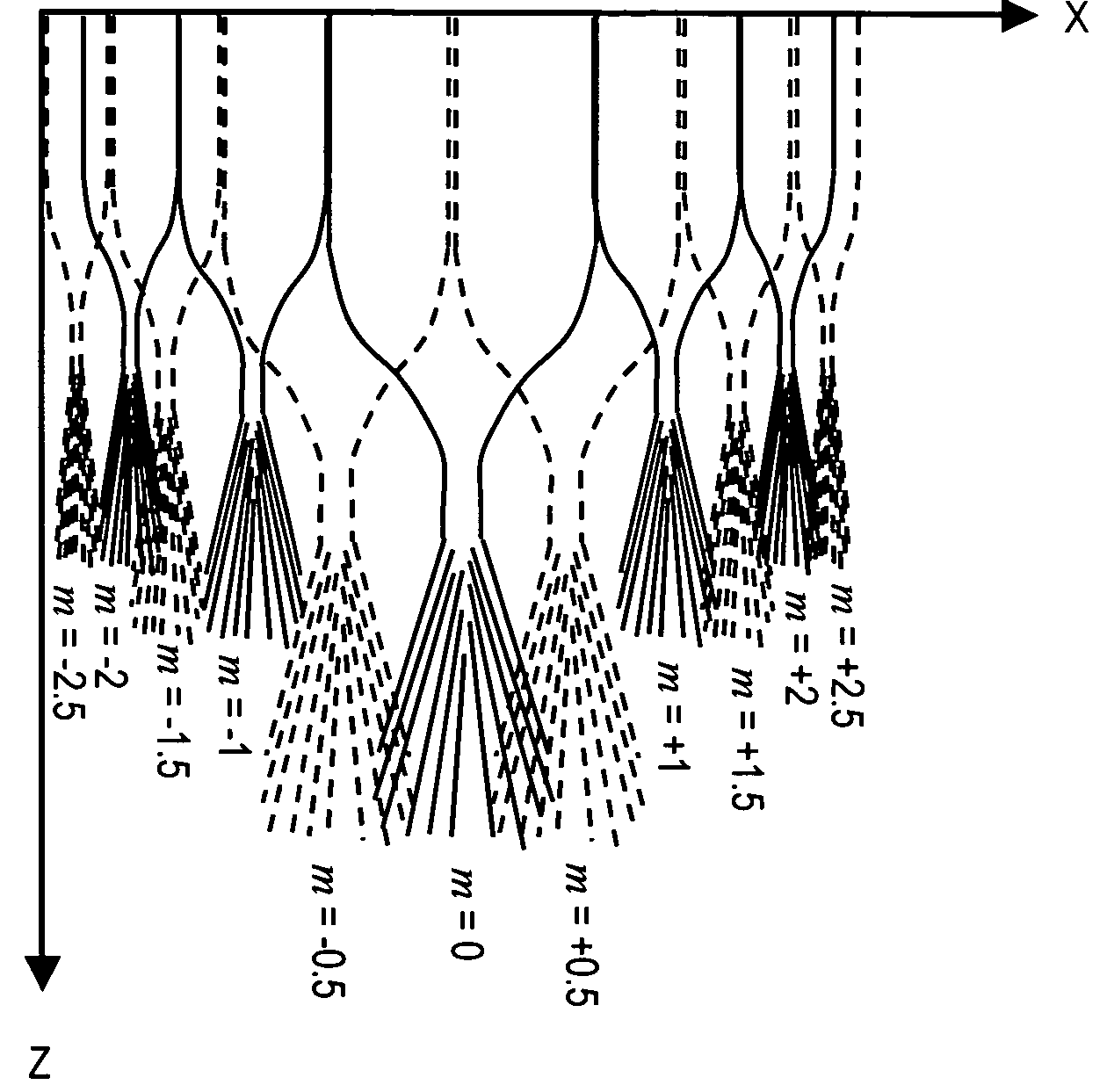
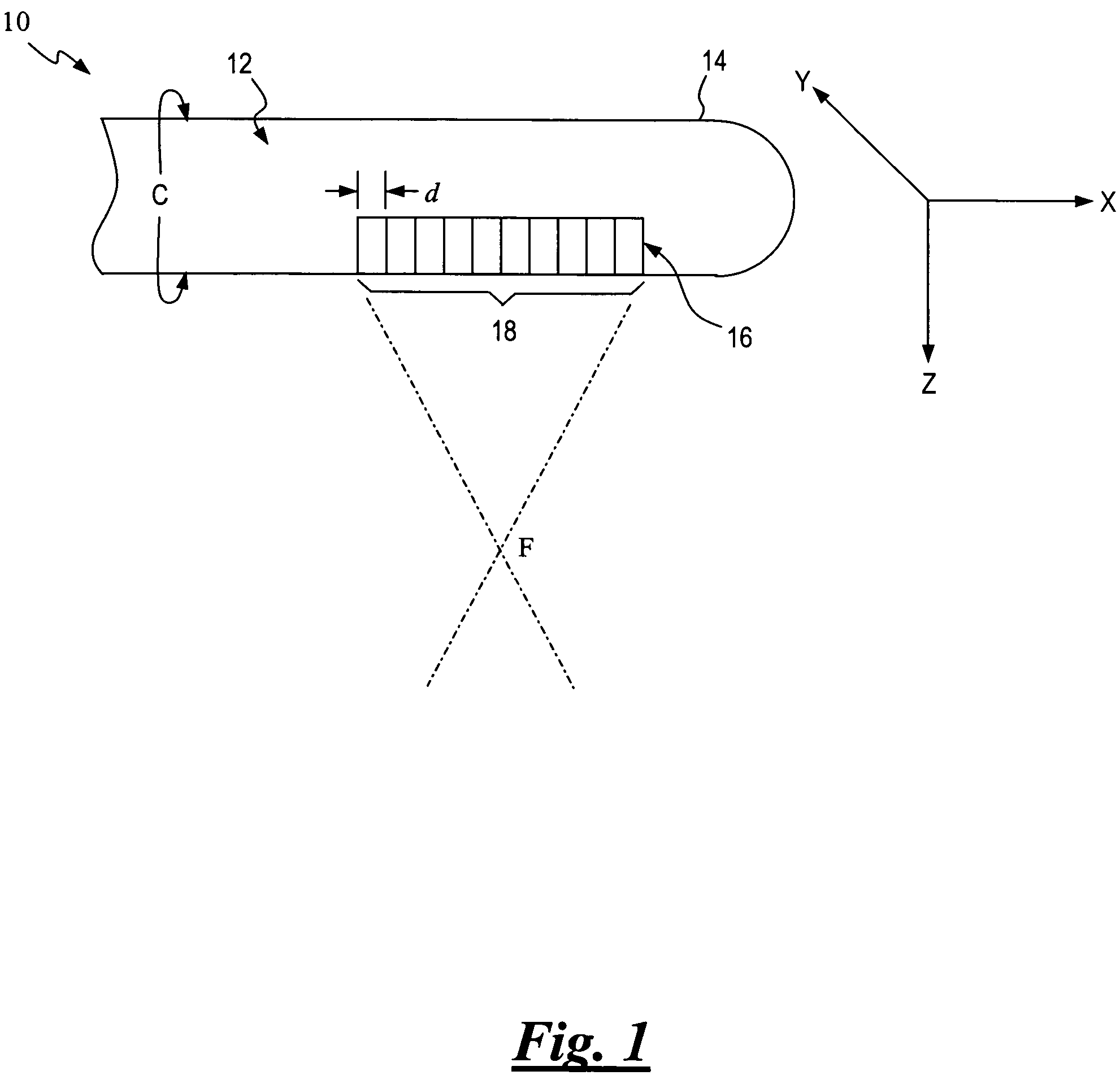
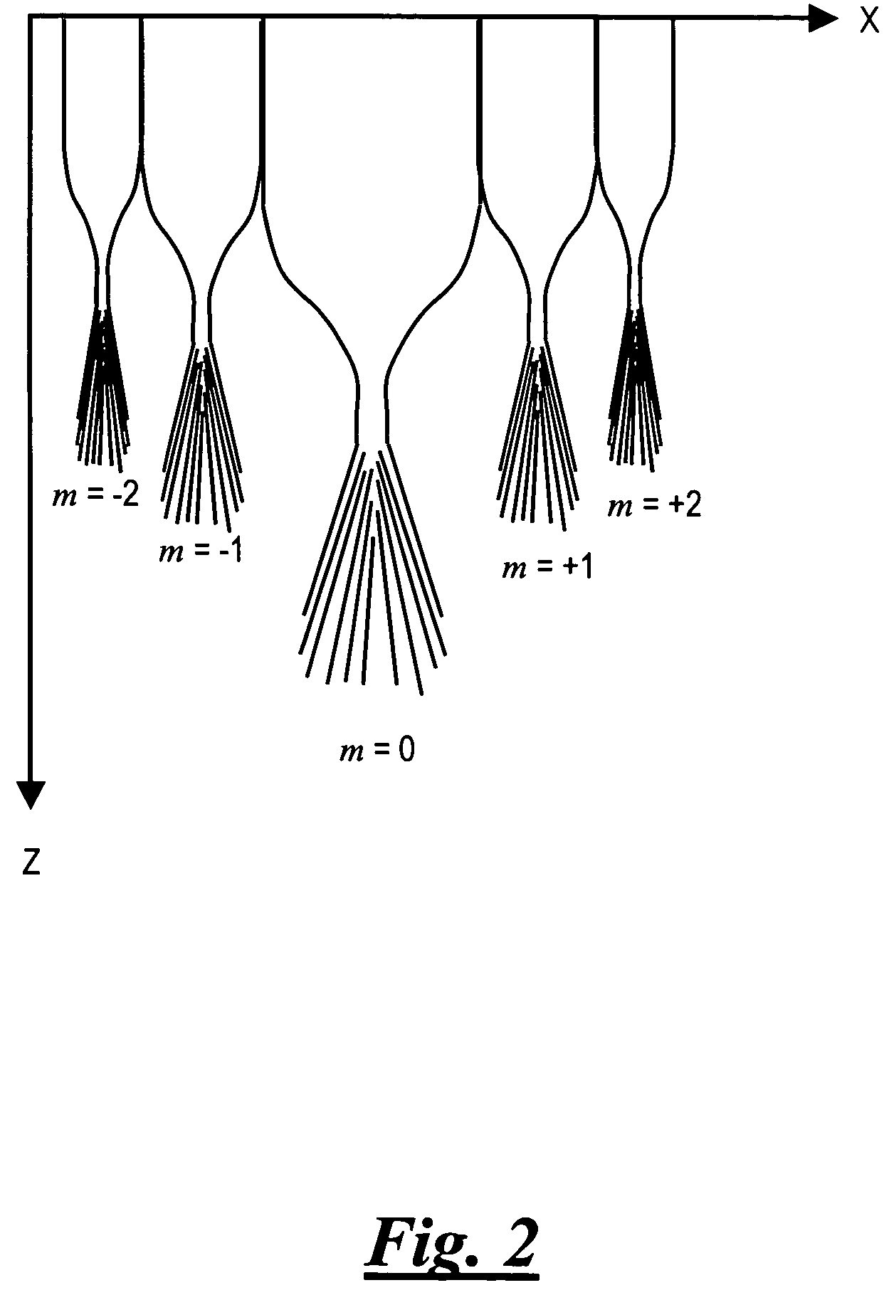
[0020]Referring now to the Figures, in which like numerals indicate like elements, FIG. 1 is a schematic view of a distal portion of an embodiment of an ultrasound treatment probe 10 that includes a body 12 in the form of a longitudinal shaft having a circumference C. Body 12 has a distal end region 14 which includes a substantially linear ultrasonic transducer array 16 having a plurality of transducer elements 18 that produce acoustic ultrasound signals used to medically treat tissue.

[0021]Probe 10 may have various configurations for various uses. For example, probe 10 may be used for laparoscopic, percutaneous and interstitial use for tissue ablation. The particular shape of probe 10 will be dictated by its use and FIG. 1 is merely intended to generally represent the distal end portion of probe 10, which typically is a cylindrical shaft. One skilled in the art will recognize that the present invention is not limited to such a configuration and may be applied to other types of arra...


PUM


Abstract

A system and method for medical treatment of tissue using ultrasound. The system comprises a probe having an array of transducer elements, an ultrasound waveform generator adapted to generate at least one electrical ultrasound signal, and a plurality of phase controls, each coupled to the ultrasound waveform generator and adapted to generate from the electrical ultrasound signal a phase-shifted drive signal that is coupled to an associated transducer element. The drive signal is effective to control grating lobe foci emitted by the array. The method employs the system.

Description

FIELD OF THE INVENTION[0001]The present invention relates generally to ultrasound, and more particularly, to an apparatus and method for providing medical treatment using high-intensity focused ultrasound.BACKGROUND OF THE INVENTION[0002]Sound waves that have a frequency greater than approximately 20 kHz are referred to in the art as “ultrasound.” In the medical field, ultrasound waves are useful for both diagnostic and therapeutic applications. Medical diagnostic ultrasound systems are useful for generating images of anatomical structures within a target area of a patient's body. The images are obtained by scanning a target area with waves of ultrasound energy. In therapeutic ultrasound applications, high intensity ultrasound energy is transmitted into a target area to induce changes in state of the target. High-intensity focused ultrasound (“HIFU”) pulses induce changes in tissue state through thermal effects (e.g., induced hyperthermia) and mechanical effects (e.g., induced cavit...

Claims


Application Information

Patent Timeline
Patent Type & Authority Patents(United States)
IPC IPC(8): A61H1/00A61H5/00A61H1/02A61N7/00A61N7/02
CPCA61N7/022A61N2007/0065A61N2007/027A61N2007/0095A61N2007/0078
Inventor MAST, T. DOUGLASFAIDI, WASEEMMAKIN, INDER RAJ S.BARTHE, PETER G.SLAYTON, MICHAEL H.
Owner CILAG GMBH INT
Why Eureka
Try Eureka