Radioactive emission detector equipped with a position tracking system and utilization thereof with medical systems and in medical procedures

a position tracking and radioactive emission technology, applied in the direction of speed measurement using gyroscopic effects, radiotherapy, sensors, etc., can solve the problems of affecting the method and outcome of surgical procedures, and reducing the accuracy of the position tracking system

Active Publication Date: 2010-11-02
SPECTRUM DYNAMICS MEDICAL LTD
View PDF171 Cites 90 Cited by
  • Summary
  • Abstract
  • Description
  • Claims
  • Application Information

AI Technical Summary

Problems solved by technology

The use of minimally invasive surgical techniques has dramatically affected the methods and outcomes of surgical procedures.
Physically cutting through tissue and organs to visually expose surgical sites in conventional “open surgical” procedures causes tremendous blunt trauma and blood loss.
Exposure of internal tissues and organs in this manner also dramatically increases the risk of infection.
The result of such open surgical procedures is more pain and suffering, higher procedural costs, and greater risk of adverse outcomes.
However, there still exist a host of technical hurdles that limit the efficacy and increase the difficulty of minimally invasive procedures, some of which were overcame by the development of sophisticated imaging techniques.
The main limitation of this system is that once the body is penetrated, scanning capabilities are limited to a translation movement along the line of penetration.
An effective collimator for gamma radiation must be several mm in thickness and therefore an effective collimator for gamma radiation cannot be engaged with a fine surgical instrument such as a surgical needle.
Thus, the system described in U.S. Pat. No. 5,846,513 cannot efficiently employ gamma detection because directionality will to a great extent be lost and cannot efficiently employ beta radiation because too high proximity to the radiative source is required, whereas body tissue limits the degree of maneuvering the instrument.
However some of these metastasis or even early stage primary tumors do not show with the imaging tools described above.
Moreover often enough the most important part of a tumor to be biopsed or surgically removed is the active, i.e., growing part, whereas using only conventional imaging cannot distinguish this specific part of a tumor from other parts thereof and / or adjacent non affected tissue.
However, in the current state of the prior art this goal is yet to be achieved, most of the common imaging modalities such as fluoroscopy, CT, MRI, mammography or ultrasound demonstrate the position and appearance of the entire lesion with anatomical modifications that the lesion causes to it's surrounding tissue, without differentiating between the non-active mass from the physiologically active part thereof.

Method used


Image

  • Radioactive emission detector equipped with a position tracking system and utilization thereof with medical systems and in medical procedures
  • Radioactive emission detector equipped with a position tracking system and utilization thereof with medical systems and in medical procedures
  • Radioactive emission detector equipped with a position tracking system and utilization thereof with medical systems and in medical procedures

Examples


Embodiment Construction

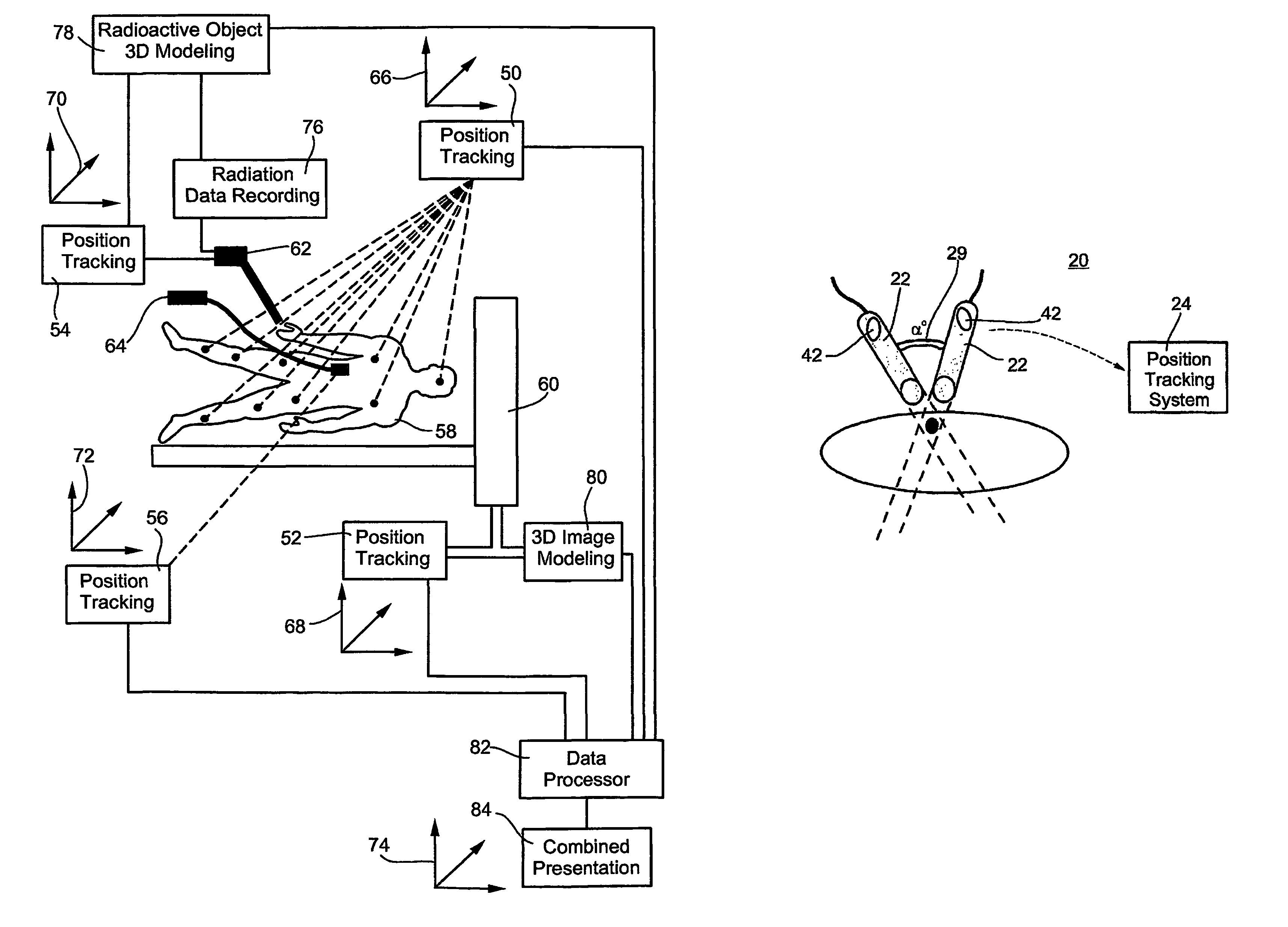
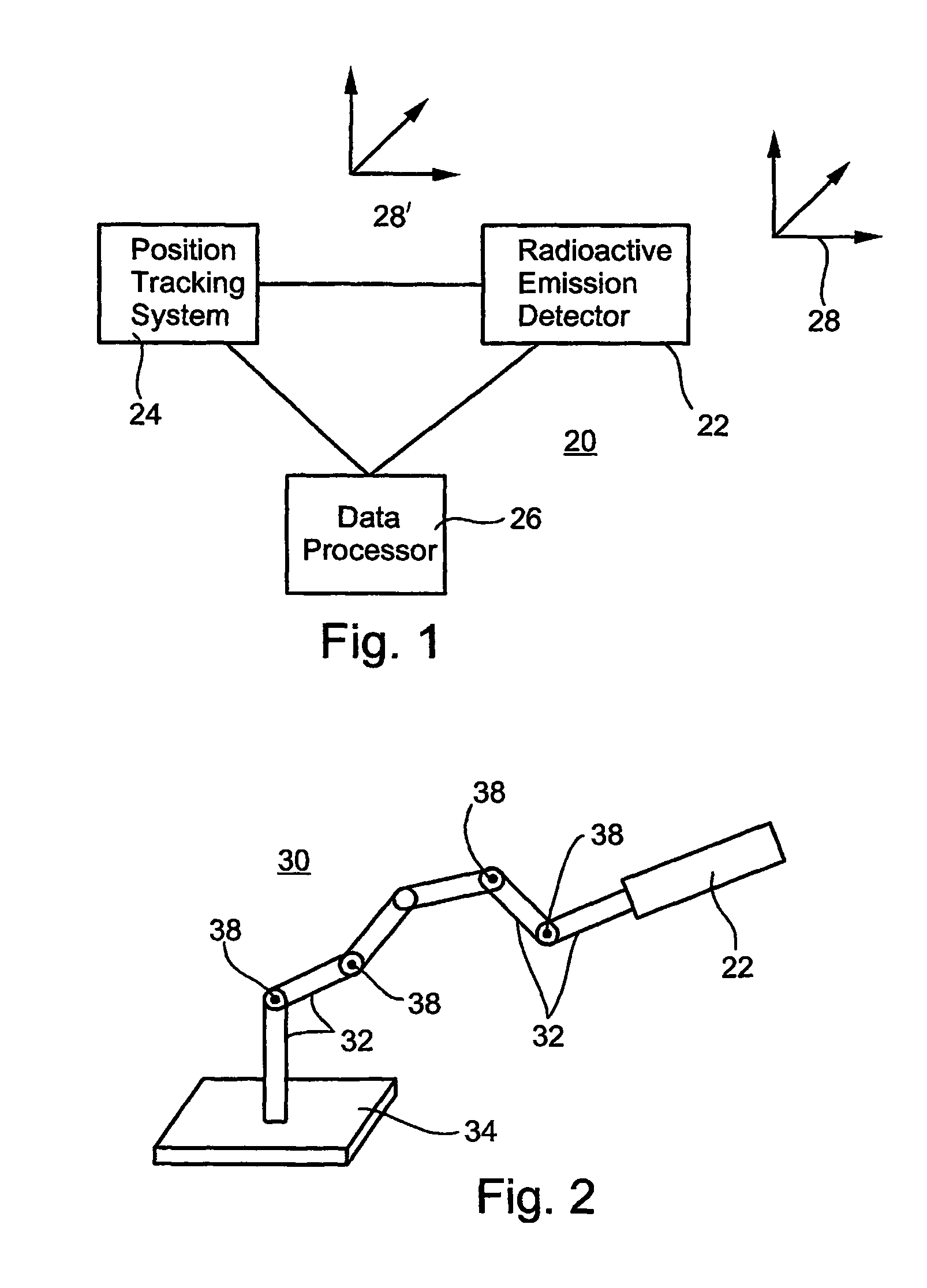
[0072]The present invention is of a radioactive emission detector equipped with a position tracking system which can be functionally integrated with medical three-dimensional imaging modalities and / or with guided minimal-invasive or other surgical tools. The present invention can be used for calculating the position of a concentrated radiopharmaceutical in the body in positional context of imaged portions of the body, which information can be used, for example, for performing an efficient and highly accurate minimally invasive surgical procedure.

[0073]The principles and operation of the present invention may be better understood with reference to the drawings and accompanying descriptions.

[0074]Before explaining at least one embodiment of the invention in detail, it is to be understood that the invention is not limited in its application to the details of construction and the arrangement of the components set forth in the following description or illustrated in the drawings. The inv...


PUM


Abstract

a system for calculating a position of a radioactivity emitting source in a system-of-coordinates, the system comprising (a) a radioactive emission detector; (b) a position tracking system being connected to and / or communicating with the radioactive emission detector; and (c) a data processor being designed and configured for receiving data inputs from the position tracking system and from the radioactive emission detector and for calculating the position of the radioactivity emitting source in the system-of-coordinates.

Description

[0001]This is a continuation-in-part of U.S. patent application Ser. No. 09 / 714,164, filed Nov. 17, 2000 now abandoned, which is a continuation-in-part of U.S. patent application Ser. No. 09 / 641,973, filed Aug. 21, 2000.FIELD AND BACKGROUND OF THE INVENTION[0002]The present invention relates to a radioactive emission detector equipped with a position tracking system. More particularly, the present invention relates to the functional integration of a radioactive emission detector equipped with a position tracking system as above with medical three-dimensional imaging modalities and / or with guided minimal-invasive surgical instruments. The present invention is therefore useful for calculating the position of a concentrated radiopharmaceutical in the body in positional context of imaged portions of the body, which information can be used, for example, for performing an efficient minimally invasive surgical procedure. The present invention further relates to a surgical instrument equipp...

Claims


Application Information

Patent Timeline
Patent Type & Authority Patents(United States)
IPC IPC(8): A61B6/00G01C19/00G06F15/00
CPCA61B19/5244A61B19/54A61B6/4258A61B19/5212A61B2017/00079A61B2019/5251A61B2019/5255A61B2019/5276A61B2019/542A61B2019/505A61B2034/2055A61B2090/392A61B34/20A61B90/361A61B2034/105A61B2034/2051A61B2090/378A61B90/39
Inventor DAVID, GAL BENZILBERSTIEN, YOELKIMCHY, YOAVAMRANI, RONI
Owner SPECTRUM DYNAMICS MEDICAL LTD
Why Eureka
Try Eureka