Method and apparatus for augmentation of a sphincter

a sphincter and sphincter technology, applied in the field of medical devices, can solve the problems of tissue fold loss, suture breakage or gradual erode through the tissue, and surgical staple loss, etc., to achieve the effect of reducing the amount of metal, avoiding image distortion, and increasing complian

Active Publication Date: 2010-11-16
COOK MEDICAL TECH LLC +1
View PDF12 Cites 17 Cited by
  • Summary
  • Abstract
  • Description
  • Claims
  • Application Information

AI Technical Summary

Benefits of technology

[0008]The foregoing problems are solved and a technical advance is achieved in an illustrative apparatus for augmenting the lower esophageal sphincter (LES) and increasing compliance, the apparatus comprising one or more bulking members that are implanted into the submucosal layer of the LES via a delivery apparatus while is a first shape or configuration, whereby they assume a second, more curvilinear configuration upon deployment. The ability of the implantable member to assume the second configuration is due to the material properties thereof. Possible materials include using a metal, polymer, biomaterial, or composite thereof capable of holding a shape memory, such as certain grades of stainless steel; a superelastic or pseudoelastic material or alloy such as nitinol; or a plastically deformable material (e.g., malleable-grade stainless steel) that would allow the implantable member to deformed into an expanded shape as it is deployed, either as it is forced into contact with tissue within the implantation site, or by the assistance of a deforming member, such as a pusher disposed within the passageway of introducer member. The implantable members can comprise materials of varying radiopacity, depending on requirements for radiographic imageability. For example, to provide compatability with CT imaging, the present implantable member may be comprised of less radiopaque (or non-radiopaque) materials to avoid image distortion or reduce the amount of metal to the minimum required to ensure adequate shape memory or resiliency. In addition, the implantable member may advantageously comprise a distinct coloration or include visually contrasting striping or portions to enhance endoscopic visualization during placement.
[0009]In a first aspect of the present invention, the bulking members comprise coiled members, such as those similar to embolization coils, which assume a regular or irregular coiled configuration upon deployment that expands within the pocket created in the submucosa and expands against adjacent tissue such that a bulge is formed about the implantation site. The implantation of the bulking member to augment LES at this site, especially when in combination with additional implanted bulking members, increases the compliance of the sphincter. The bulking members may comprise any suitable material such as a stainless steel, or shape memory metal, alloy, or polymer (e.g., nitinol) and may further comprise attached fibers, outer polymer or biomaterial layers or coating, and / or pharmacologically active substances applied to or incorporated therewithin.
[0011]In another aspect of the invention, the implantable member comprises an anchoring point for sutures during a gastroplication procedure to create folds at the LES to increase compliance. One of the disadvantages of gastroplication of the LES using sutures is that over time, the sutures may break or gradually erode through the tissue and pull free. When this occurs, the tissue fold created by the suturing procedure may be lost. To help prevent the sutures used to create the folds from being pulled free over time, the sutures may be looped around or through a single or combination of bulking / anchoring members or they may be tied or otherwise secured directly thereto. Alternatively, the bulking / anchoring members can be configured to serve as anchor points for other means of creating gastroplication folds, such as clips, staples, rings, etc.

Problems solved by technology

One of the disadvantages of gastroplication of the LES using sutures is that over time, the sutures may break or gradually erode through the tissue and pull free.
When this occurs, the tissue fold created by the suturing procedure may be lost.

Method used


Image

  • Method and apparatus for augmentation of a sphincter
  • Method and apparatus for augmentation of a sphincter
  • Method and apparatus for augmentation of a sphincter

Examples


Embodiment Construction

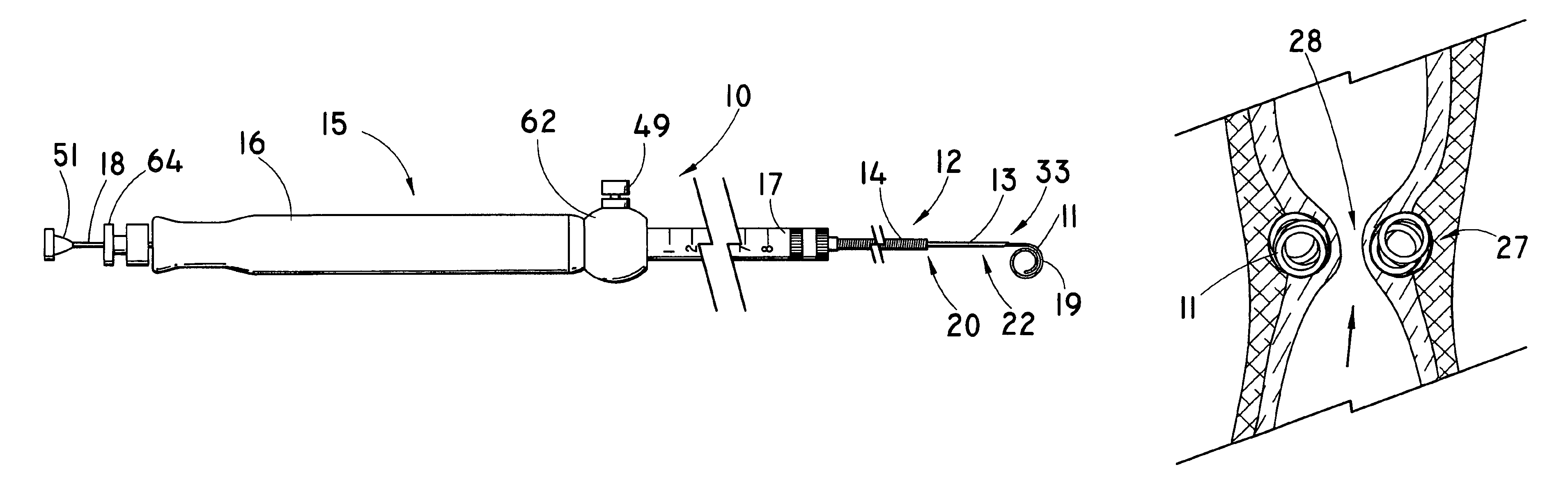
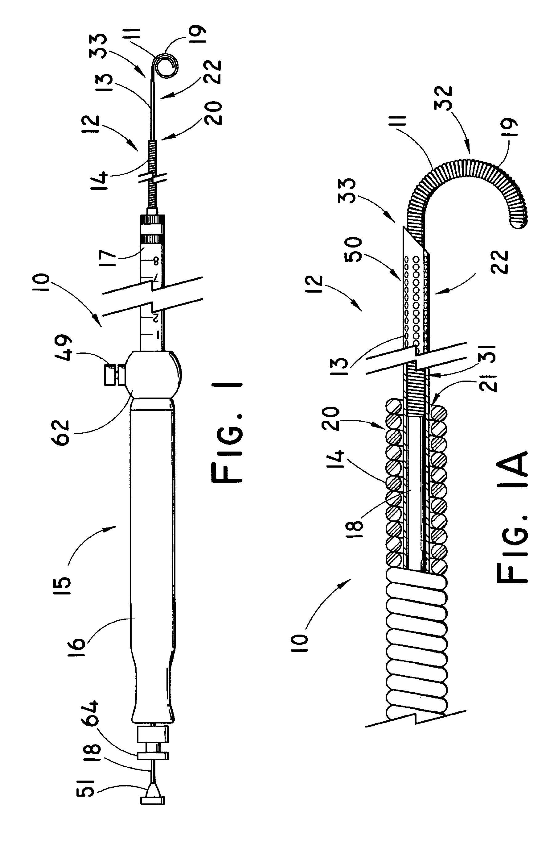
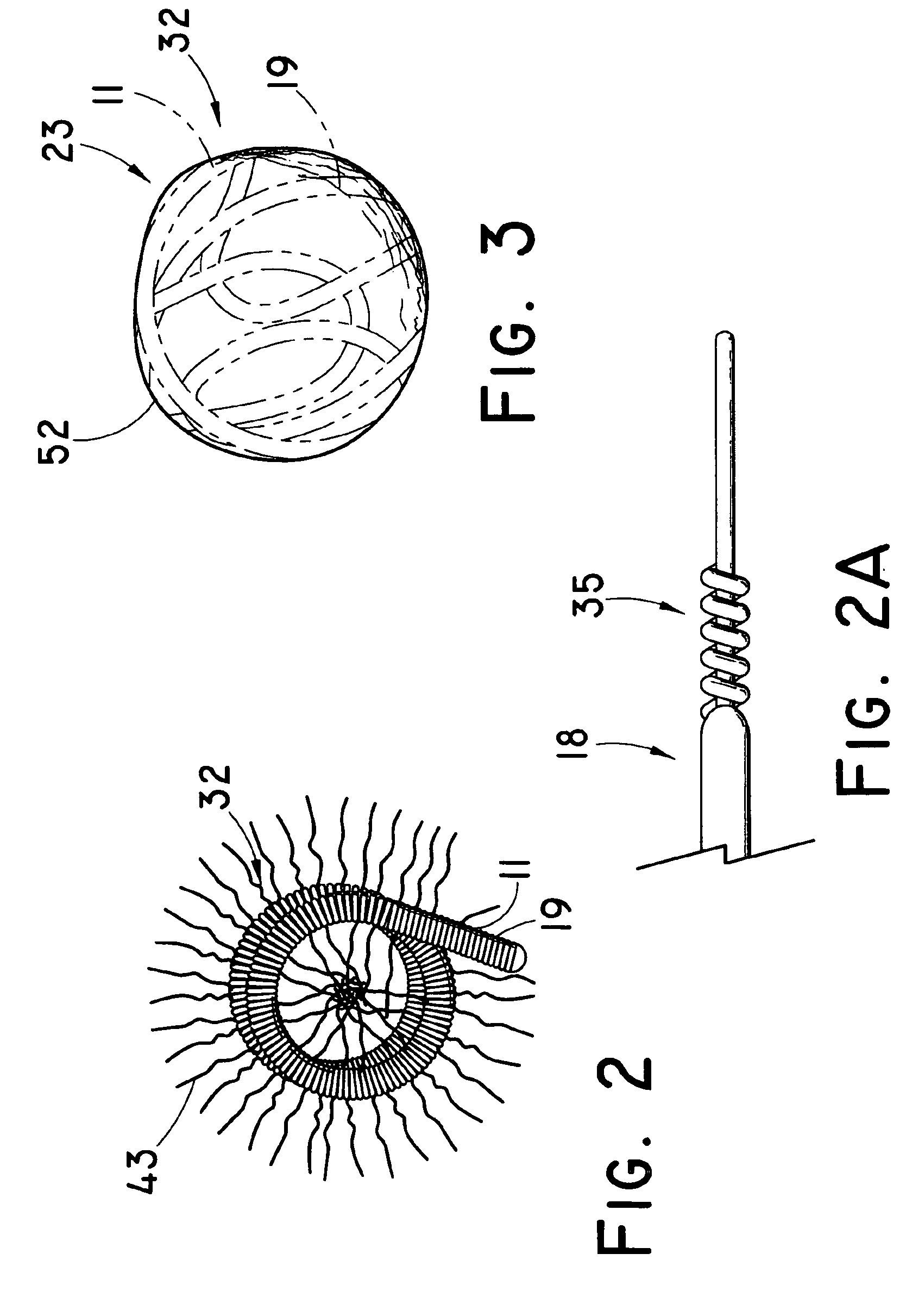
[0031]The present invention, exemplary embodiments of which are depicted in FIGS. 1-18, includes an apparatus 10 and method for introducing one or more implantable members 11 using an introducer member 13, such as an endoscopic needle, into a pocket formed within the submucosal layer of the gastroesophageal (GE) junction or lower esophageal sphincter (LES) 27 to augment and tighten the LES to provide a more effective barrier against stomach acid reflux. In the illustrative embodiment depicted in FIGS. 1 and 1A, the apparatus 10 comprises a delivery system 12 that is similar in configuration to the ECHOTIP® Ultrasound Needle (Wilson-Cook Medical, Inc.), which includes a needle portion 33 (e.g., 19 ga), typically having a beveled distal tip 33, that is extendable from an outer sheath portion 14 (comprised of a coiled sheath member in this particular embodiment), each of which are attached to a coaxial handle assembly 15. The coaxial handle assembly 15 comprises a first portion 16 atta...


PUM


Abstract

A system and method for delivering one or more implantable members, such as metal coils, via an introducer member, such as an endoscopic needle, into a space created within the submucosal layers of the LES to augment and bulk the sphincter to improve compliance in patients with gastroesophageal reflux disease. The implantable member comprises a first substantially straightened configuration when carried by the introducer member, whereby it assumes a more curvilinear or coiled configuration following deployment such that it expands against adjacent submucosal tissue, creating a bulge at the implantation site. In another embodiment, the implantable member can serve as an anchoring port for sutures or other elements introduced for gastroplication of the LES to create tissue folds that increase sphincter compliance. The implantable members may be configured to include an external portion that interconnects with an adjacent implantable member, thereby creating tension that also increases compliance of the LES.

Description

CROSS-REFERENCE TO RELATED APPLICATIONS[0001]This application claims priority of provisional application Ser. No. 60 / 636,131, filed Dec. 15, 2004.TECHNICAL FIELD[0002]This invention relates to medical devices, more particularly to implant materials used to augment the function of a sphincter, particularly those delivered endoscopically, such as by a needle.BACKGROUND OF THE INVENTION[0003]It has been estimated that 7 million Americans suffer from progressive gastroesophageal reflux disease (GERD) which is characterized by frequent episodes of heartburn, typically on at least a daily basis. Without adequate treatment, GERD can cause erosion of the esophageal lining as the lower esophageal sphincter (LES), a segment of smooth muscle located at the junction of the stomach and the esophagus, gradually loses its ability to function as the barrier that prevents stomach acid reflux.[0004]While drug therapies, such as proton pump inhibitors, have become a viable option for many of those dia...

Claims


Application Information

Patent Timeline
Patent Type & Authority Patents(United States)
IPC IPC(8): A61F2/04
CPCA61B17/00234A61B17/12022A61B17/12099A61B17/12145A61B17/1215A61B17/3468A61F2/0036A61B17/0401A61B2017/00867A61B2017/0649A61B2017/1205
Inventor LEHMAN, GLEN A.RUCKER, BRIAN K.
Owner COOK MEDICAL TECH LLC
Why Eureka
Try Eureka