Compact and mobile high resolution PET brain imager

a brain imaging and mobile technology, applied in the field of pet imaging, can solve the problems of large scanners, insufficient brain imaging modalities such as pet+ct and mri+pet, and high cost, and achieve the effects of not being optimized for brain imaging, not being able and being unable to meet the needs of patients

Active Publication Date: 2011-02-08
JEFFERSON SCI ASSOCS LLC
View PDF5 Cites 25 Cited by
  • Summary
  • Abstract
  • Description
  • Claims
  • Application Information

AI Technical Summary

Benefits of technology

[0007]Several advantages are achieved with the compact high resolution brain imager of the present invention, including:

Problems solved by technology

Although many new PET radiopharmaceuticals for brain function imaging are under development, imaging modalities for the brain such as PET+CT and MRI+PET are insufficient due to poor PET resolution and poor CT / MRI / fMRI specificity.
Current clinical PET scanners have a resolution of 4-5 mm, which is insufficient in many brain imaging situations.
In addition, current clinical PET scanners are large, expensive, not optimized for brain imaging, and usually are available only in a package with CT.
The existing standard PET imagers are bulky devices that are placed in dedicated imaging rooms and require patient to be brought to the imager.
Some patients cannot be transported to the imaging room, which could be in a far away part of the medical complex, particularly in situations when they need to be hooked to life-saving and monitoring machines or when quick diagnostic and staging decision is important.
In the current state of the art, brain imagers are bulky and heavy devices and are not capable of providing dynamic high resolution 2D or 3D images.

Method used


Image

  • Compact and mobile high resolution PET brain imager
  • Compact and mobile high resolution PET brain imager
  • Compact and mobile high resolution PET brain imager

Examples


first embodiment

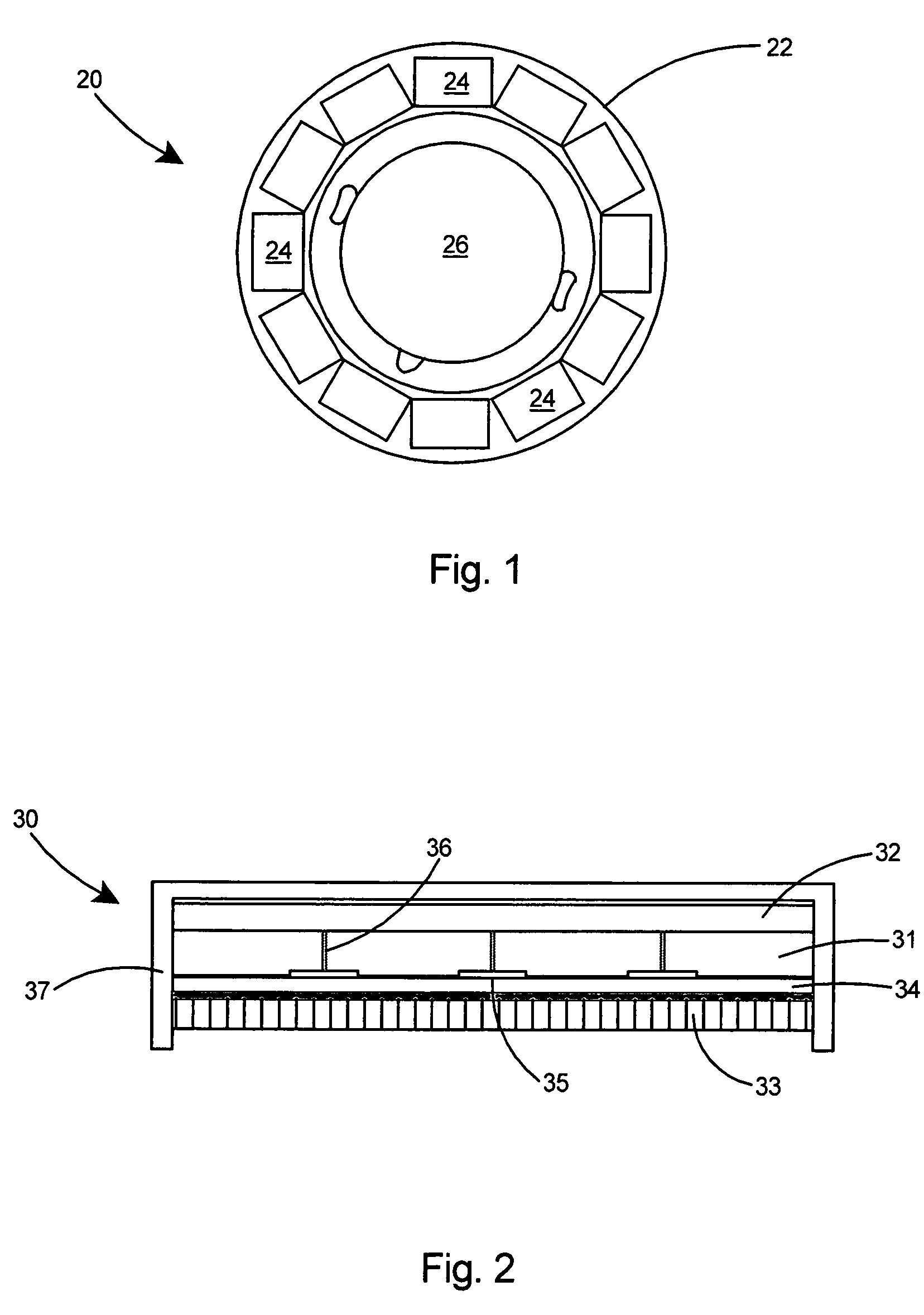
[0078]Referring to FIG. 2 there is shown a PET photodetector module 30 for constructing the compact brain imager of the present invention. The photodetector module 30 is based on compact, 1″ or 2″ in size, position sensitive photomultipliers (PSPMTs) 31 from Hamamatsu Corporation of Bridgewater, N.J. or Burle Industries of Lancaster, Pa. The photomultipliers 31 are coupled to an array 33 of 2×2×10 mm LYSO scintillator pixels. In the present invention, a modular approach is used in constructing the PET photodetector module 30. The detector module 30 includes an array of compact H8500 or H9500 flat panel PMTs 31 using high-rate resistive readout electronics 32. The position sensitive flat panel PMTs 31 are placed in a tight array and coupled to the scintillator array 33 through a window 34 and an optical light guide. The flat panel Hamamatsu H8500 or H9500 PMTs are each approximately 5 cm×5 cm in size, to obtain coverage of about 20 cm per detector module 30. Reflective strips 35 are ...

second embodiment

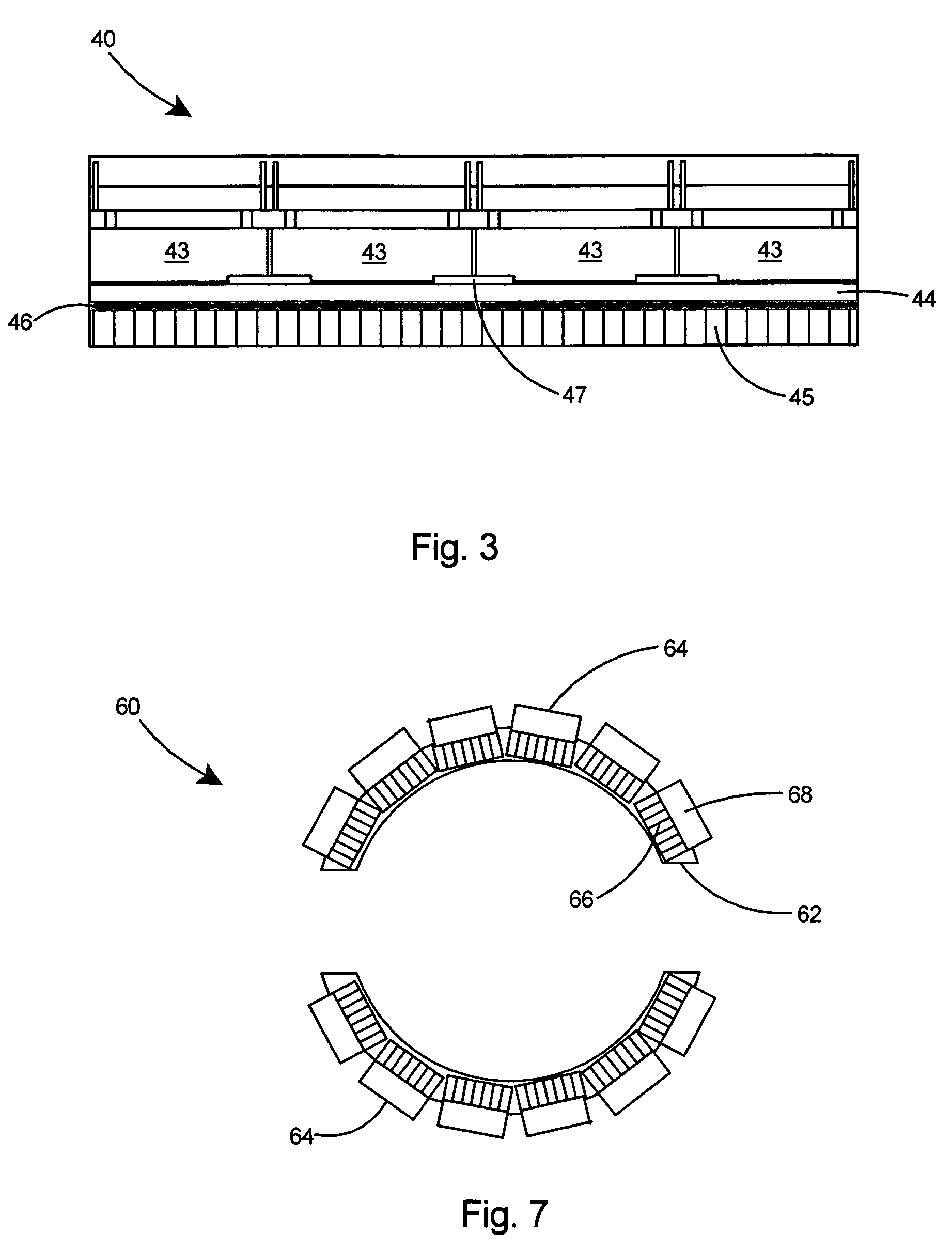
[0079]Referring to FIG. 3 there is shown a compact brain PET detector module 40 based on an array of 16 (4×4) Burle 85001-501 MCP (microchannel plates) PMTs 43 available from Burle Industries of Lancaster, Pa. The 85001-501 PMTs 43 are coupled through optical spreader window 44 to a scintillation array 45 encapsulated behind a window 46. Reflective strips 47 are placed in the dead regions between the MCP PMTs to improve scintillation light collection from ˜2 cm wide dead regions between these MCP PMTs. The shielding is omitted in this figure.

third embodiment

[0080]Referring to FIGS. 4-6, a third and preferred embodiment of the PET detector head is produced using silicon photomultipliers (SiPMTs) in lieu of the position sensitive PMTs. the photodetector module 50 is the preferred construction for the brain imager of the present invention as the SiPMTs provide a more compact and lighter weight imager. Typically SiPMT modules come in smaller size units of approximately 3 mm×3 mm to 5 mm×5 mm area and 1.5 mm in thickness. When combined with onboard electronics, the thickness of the SiPMT plus onboard electronics is less than 1 cm. Arrays of SiPMTs are needed to cover the desired active field of view. FIGS. 4-6 show an example of how to achieve a silicon PMT based photodetector 50 (FIG. 6) of approximately 5 cm×5 cm active field of view using nominal 12.5 mm silicon PMT basic imaging modules 52 (FIG. 4) each composed of sixteen 3 mm basic silicon PMT units 54. As shown in FIG. 4, the basic initial imaging module 52 can have an array of sixte...


PUM


Abstract

A brain imager includes a compact ring-like static PET imager mounted in a helmet-like structure. When attached to a patient's head, the helmet-like brain imager maintains the relative head-to-imager geometry fixed through the whole imaging procedure. The brain imaging helmet contains radiation sensors and minimal front-end electronics. A flexible mechanical suspension / harness system supports the weight of the helmet thereby allowing for patient to have limited movements of the head during imaging scans. The compact ring-like PET imager enables very high resolution imaging of neurological brain functions, cancer, and effects of trauma using a rather simple mobile scanner with limited space needs for use and storage.

Description

[0001]The United States of America may have certain rights to this invention under Management and Operating contract No. DE-AC05-06OR23177 from the Department of Energy.FIELD OF THE INVENTION[0002]This invention relates to PET imaging and more particularly to a compact and mobile high resolution PET imaging system for imaging the neurological brain functions of a patient.BACKGROUND OF THE INVENTION[0003]Brain diseases, such as Alzheimer's and Parkinson's, are becoming more prevalent in the aging population because of increased life span. As US and European populations continue to age, Alzheimer's disease will increase with the US expected to have 16 million cases by 2050. An estimated 1.5 million people in the US have Parkinson's, which affects 1% of Americans over 60 and includes about 100,000 new cases each year.[0004]Although many new PET radiopharmaceuticals for brain function imaging are under development, imaging modalities for the brain such as PET+CT and MRI+PET are insuffic...

Claims


Application Information

Patent Timeline
Patent Type & Authority Patents(United States)
IPC IPC(8): G01T1/161
CPCA61B6/037G01T1/00
Inventor MAJEWSKI, STANISLAWPROFFITT, JAMES
Owner JEFFERSON SCI ASSOCS LLC
Why Eureka
Try Eureka