Atraumatic tissue retraction device

a tissue retraction and atraumatic technology, applied in the field of atraumatic tissue retraction devices, can solve the problems of significant physical trauma to the patient, require one week of hospital recovery time, and require weeks of convalescence, so as to improve the access to the surgical site, improve the surgical effect, and adjust the tensioning of the straps

Active Publication Date: 2011-03-22
ATRICURE
View PDF9 Cites 40 Cited by
  • Summary
  • Abstract
  • Description
  • Claims
  • Application Information

AI Technical Summary

Benefits of technology

[0013]The flexible straps of the surgical retractor may be constructed of a soft, resilient material, such as silicone, which provide an atraumatic barrier between the ribs and soft tissues adjacent to the incision site. This reduces the amount of trauma to the ribs and soft tissues caused by various surgical instruments (ie. rib spreaders, surgical tools, etc)
[0027]The present invention can be used to provide tissue retraction and expand access openings by means of the strength and elastic properties of the materials used to manufacture the retractor. The present invention is also configured to be couplable with an adjustable surgical retractor with blades to be inserted into the patient cavity for large expansion against opposing thoracic structures while providing atraumatic contact with those anatomical structures.
[0028]The present invention provides the ability to function to retract tissues, without significant expansion of the incision, but also to enlarge the size of the incision, a function that is provided by the tear strength of the silicone or the ability to combine it with an adjustable surgical blade.
[0029]The present invention provides the ability to adjust the tensioning of the straps even after initial deployment through the use of the hook and loop material. This may be necessary when a surgeon needs to enlarge the incision to gain better access to the surgical site. Competitive devices use an adhesive that does not allow for repositioning subsequent to initial deployment.

Problems solved by technology

This technique causes significant physical trauma to the patient and can require one week of hospital recovery time and up to eight weeks of convalescence.
This can be very expensive in terms of hospital costs and disability, to say nothing of the pain to the patient.
While this procedure has the advantage of being less invasive or traumatic than performing a media, sternotomy, there are numerous disadvantages to using trocars to establish the entry ports for the instruments and viewscope.
In addition, because of the limited movement, the surgeon often has to force the instrument into an angle that moves the trocar and further damages the surrounding tissue and nerves.
The need to force the instrument causes the surgeon to lose sensitivity and tactile feedback, thus making the surgery more difficult.
Major disadvantages of these systems include their limited positioning, complexity, and trauma to the surrounding tissue.

Method used


Image

  • Atraumatic tissue retraction device
  • Atraumatic tissue retraction device
  • Atraumatic tissue retraction device

Examples


Embodiment Construction

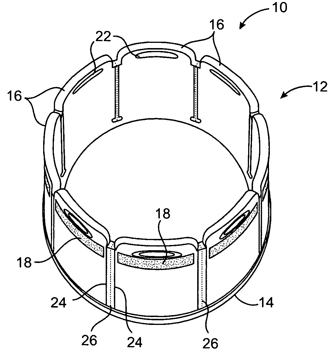
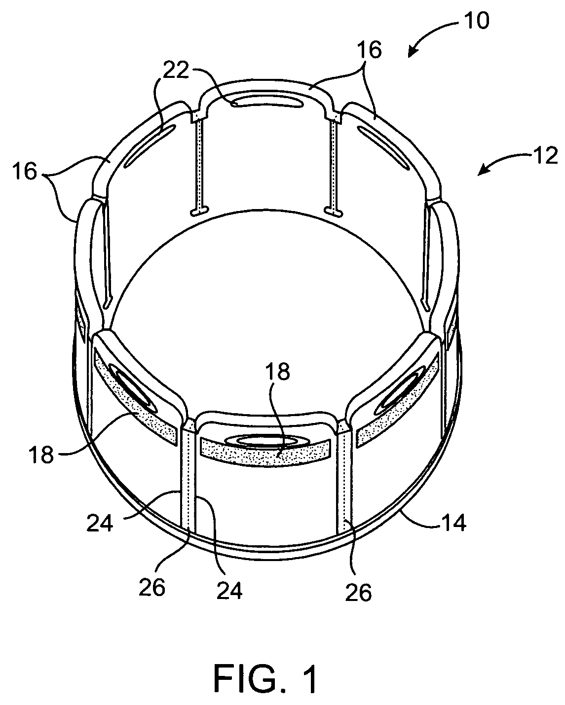
[0038]FIG. 1 is a top perspective view of the atraumatic tissue retraction device 10. The atraumatic tissue retractor 10 is a device used to keep the field of view of a thoracotomy, sternotomy, or other surgical portal clear of soft tissues, as well as limit or prevent trauma to the ribs and other soft tissues. The device includes a sleeve 12 with an elastic or flexible ring 14 in the lower or distal end. The ring 14 may be formed of any resilient or elastic material, such as Nitinol, other metals, and plastics. The ring 14 may be adhered, bonded, overmolded into or otherwise connected to the base of the sleeve 12. The sleeve 12 is divided into a multiplicity of straps 16. Any suitable number of straps 16 may be used, preferably from three to twenty, more preferably in the range of four to fourteen and most preferably from six to ten. In the embodiment shown, eight straps 16 are used. Any soft resilient, flexible material may be used to form the sleeve. One suitable material is sili...


PUM


Abstract

Methods and apparatus for a surgical retractor include a ring, a plurality of flexible straps connected to the ring, a patch of hook or loop material connected to each strap, a coordinating patch of hook or loop material connectable to the patient's skin or the surgical drape. The flexible straps of the surgical retractor may be frangibly connected together. LEDs molded into the distal end create a light source to illuminate the surgical site. The ring may take several forms including a flexible or adjustable ring and an inflatable bladder. The ring of the surgical retractor is inserted into the surgical incision, a patch of loop fastener is attached to the patient, a set of straps connected to the ring are pulled outward and the hook portion is applied to the loop portion to hold the incision open. The retractor is useable for thoracic and other types of surgery.

Description

FIELD OF THE INVENTION[0001]The present invention pertains to apparatus and methods for atraumatically retracting tissue. In particular, the present invention creates and maintains an opening through soft tissue.BACKGROUND OF THE INVENTION[0002]Surgery on the heart is one of the most commonly performed types of surgery that is done in hospitals across the U.S. Cardiac surgery can involve the correction of defects in the valves of the heart, defects to the veins or the arteries of the heart and defects such as aneurysms and thromboses that relate to the circulation of blood from the heart to the body. In the past, most cardiac surgery was performed as open-chest surgery, in which a primary median sternotomy was performed. That procedure involves vertical midline skin incision from just below the super sternal notch to a point one to three centimeters below the tip of the xiphoid. This is followed by scoring the sternum with a cautery, then dividing the sternum down the midline and sp...

Claims


Application Information

Patent Timeline
Patent Type & Authority Patents(United States)
IPC IPC(8): A61B1/32
CPCA61B1/32A61B17/02A61B17/3423A61B17/3439A61B1/0607A61B1/0676A61B1/0684A61B2017/0287
Inventor BANCHIERI, MIKEIBRAHIM, TAMERBERTOLERO, RAYMOND
Owner ATRICURE
Why Eureka
Try Eureka