Anatomical marker for x-ray orientation

a technology of anatomical markers and x-rays, applied in optics, instruments, electrical equipment, etc., can solve the problems of difficult to ascertain the body position of a patient in a given fluoroscopy image, difficult to quickly, clearly, and confidently delineate left, right, and cranio-caudal positions, and markers in the related art consistently do not provide simultaneous orientation as to antero-posterior and cranio

Inactive Publication Date: 2011-07-12
MDX LAB
View PDF30 Cites 37 Cited by
  • Summary
  • Abstract
  • Description
  • Claims
  • Application Information

AI Technical Summary

Benefits of technology

[0013]It is an object of the present invention to reduce error from a misoriented x-ray image that would otherwise result from a conventional left or right marker. It is another object of the invention to reduce error from a misoriented x-ray image that results from ambiguity as to left, right, cranial, caudal, anterior or posterior orientation of a patient's body position. It is still another object of the present invention to quickly and effectively orient the patient's left, right, cranial, and caudal directions in a localized fluoroscopy image with a single-piece marker.

Problems solved by technology

One problem is that images must be oriented in left-right, cranio-caudal, and antero-posterior position before a procedure can begin.
As a result, it is difficult to ascertain the body position of a patient in a given fluoroscopy image.
Also, given the axial symmetry of bony landmarks in x-ray, it is difficult to quickly, clearly, and confidently delineate left, right, and cranio-caudal positions.
However, markers in the related art consistently do not provide simultaneous orientation as to antero-posterior, cranio-caudal and left-right positioning by a single marker.
Also, placing an “R” marker on the patient's left side results in incorrect image orientation; the “R” appears on the right side of the image, which initially appears correct.
Misorientation can lead to accidents in medical procedures if an incorrect position is not identified, especially prior to invasive procedures.
If the incorrect position is only identified after a procedure begins, the misoriented image has to be corrected during the procedure, increasing procedure time.
Multiple individual markers could be used to provide simultaneous orientation as to multiple directions, but their relative orientation is not fixed and is subject to error.
Also, using multiple markers requires extra time, slowing down positioning.
Separate pieces can also be easily lost.
Further, the spine is not always in view.

Method used


Image

  • Anatomical marker for x-ray orientation
  • Anatomical marker for x-ray orientation
  • Anatomical marker for x-ray orientation

Examples


Embodiment Construction

[0036]In the following description of the preferred embodiments, reference is made to the accompanying drawings that form a part hereof, and in which is shown by way of illustration specific embodiments in which the invention may be practiced. It is to be understood that other embodiments may be utilized and structural changes may be made without departing from the scope of the present invention.

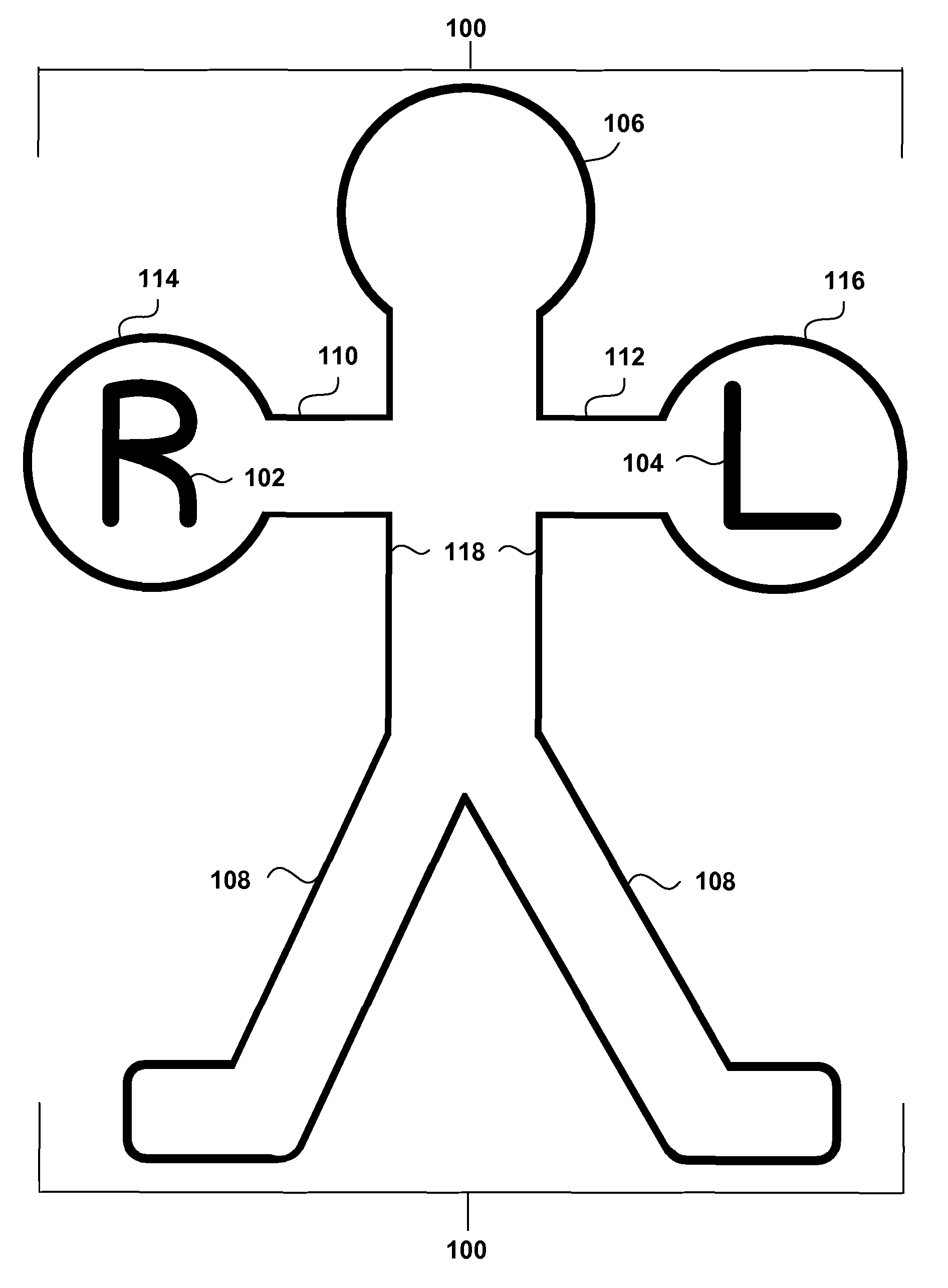
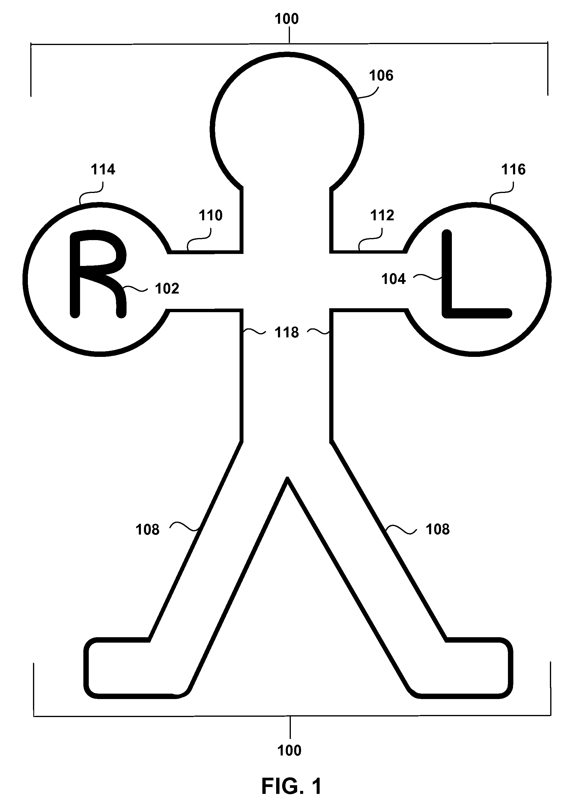
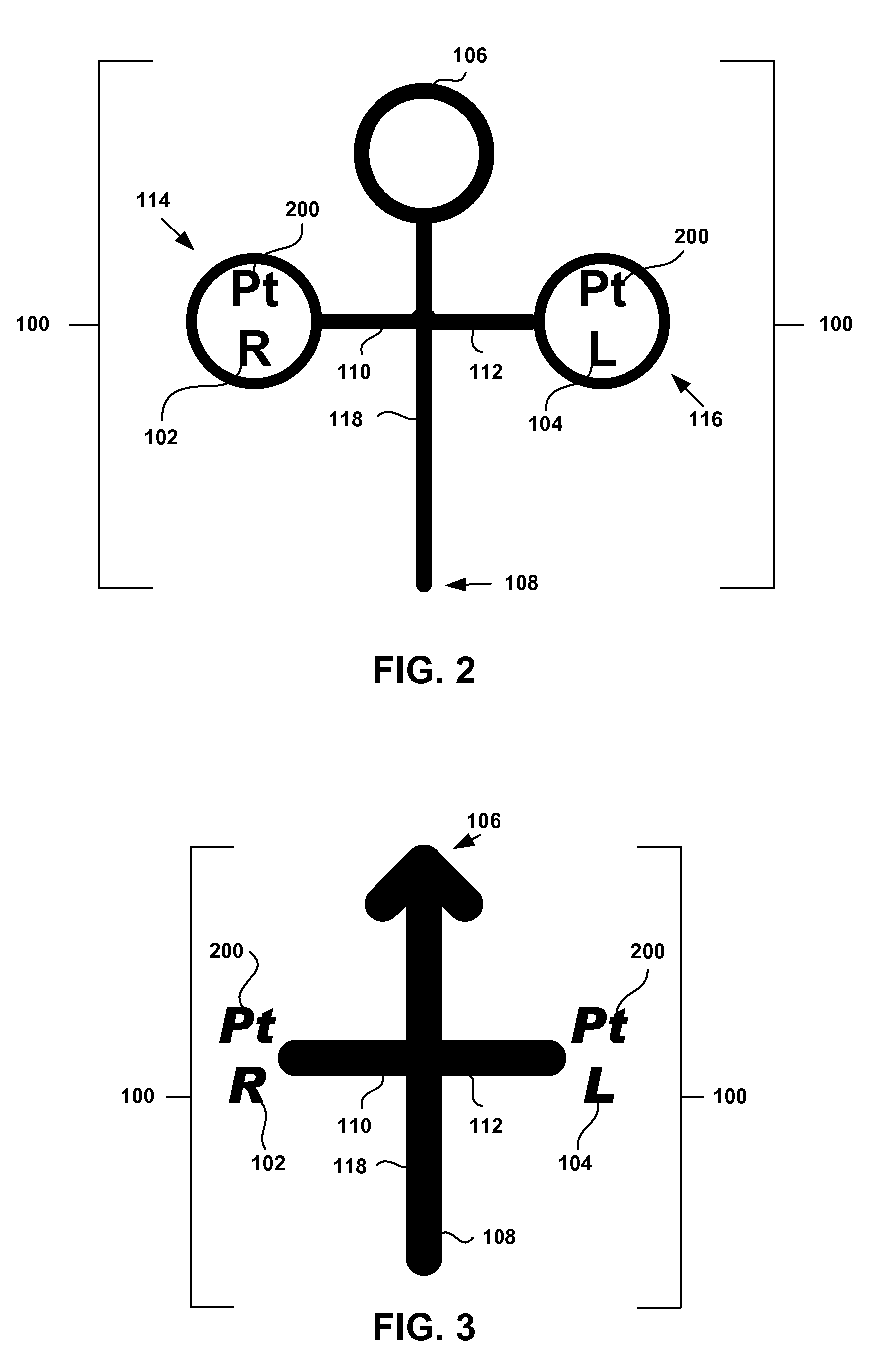
[0037]To aid in a basic understanding of a possible simplified embodiment of the present invention, and referring generally to the figures and numeric references throughout, marker 100 can be embedded in radiolucent material 400 with first side indicator 102, second side indicator 104, cranial indicator 106, caudal indicator 108, anterior indicator 600, and posterior indicator 700.

[0038]It will be understood that “x-ray” can refer to any radiological technique. It will be understood that “radiological image,”“fluoroscopy image,”“x-ray image,” and “image” can be used interchangeably and can e...


PUM


Abstract

A radiopaque marker for more effectively orienting a fluoroscopy image to a patient's body position is disclosed. A method for orienting a radiological image with a patient's anatomical position comprises the step of forming a marker with a first side indicator and a second side indicator formed substantially opposite the first side indicator. A device in accordance with the present invention comprises a radiopaque marker comprising a first side indicator, a second side indicator, and a humanoid shape.

Description

BACKGROUND OF THE INVENTION[0001]1. Field of the Invention[0002]The present invention relates in general to markers used in radiology, and in particular to an anatomical marker used to orient to a patient's anatomical position in x-ray and fluoroscopy procedures.[0003]2. Description of the Related Art[0004]Markers are often used to identify a body part shown in an x-ray image. X-rays which are absorbed by markers do not appear on x-ray sensitive media. In fluoroscopy, real-time x-ray images are displayed on a video monitor. Fluoroscopy is often combined with other medical procedures.[0005]Paramount to fluoroscopy is initially orienting the x-ray image to the patient's body position. One problem is that images must be oriented in left-right, cranio-caudal, and antero-posterior position before a procedure can begin. Although bony landmarks are often used to orient the patient's body position with the image, fluoroscopy images typically do not include the entirety of a patient's body. ...

Claims


Application Information

Patent Timeline
Patent Type & Authority Patents(United States)
IPC IPC(8): H05G1/28
CPCG03B42/047
Inventor NGO, PETER D.
Owner MDX LAB
Why Eureka
Try Eureka