Autogenic living scaffolds and living tissue matrices: methods and uses thereof

a technology of living scaffolds and living tissue, applied in the field of living scaffolds, can solve the problems of limited durability of mechanical devices, imperfect matches, tissue complications, etc., and achieve the effect of preventing host rejection and thickening and strengthening

Inactive Publication Date: 2011-10-25
GENESIS TECH LTD
View PDF29 Cites 29 Cited by
  • Summary
  • Abstract
  • Description
  • Claims
  • Application Information

AI Technical Summary

Benefits of technology

[0014]In another embodiment, the Autogenic Living Scaffold may also be grown into specific shapes by molds, and may also be reshaped to some degree. For example, a sheet of the above example of a Fibroblast Autogenic Living Scaffold, seeded first with astrocytes and then nerve cells, may be rolled into a cylinder. This cylinder may then be implanted into a spinal cord in vivo. The Autogenic Living Scaffold also provides mechanical support to the seeded cells. For example, in a particular embodiment, a Fibroblast Autogenic Living Scaffold seeded with neurons may be mechanically stressed and compressed, without major damage to the neurons, even though such a degree of mechanical stress and compression kills most neurons when grown in the absence of an ALS. The Autogenic Living Scaffold of other embodiments may also be introduced to mechanical stress or tension which may change the properties of the Living Scaffold and any cells or tissue that are growing on it.
[0015]In one particular embodiment, the fibers of a Fibroblast Autogenic Living Scaffold may also be made to grow in parallel, which helps seeded nerve cells to also grown in parallel along these fibers, especially when Schwann cells are previously seeded onto the scaffolds and first start growing in parallel along these fibers. This may be even more useful when implanted in the spinal cord, since the implanted nerve cells may then be aligned in the same direction as the native nerve cells in the spinal cord. In still another embodiment, a sheet of Autogenic Living Scaffold with the seeded neurons may also be rolled into a cylinder prior to implantation to produce a structure with layers of neurons aligned in the same general direction as the native neurons in the spinal cord. Similar things may be done for implantation into other tissues and organs.
[0018]In the case of particular embodiments of fALS of the present application, the high density of fibroblasts along with the insoluble ECM proteins collagen and fibronectin of the ALS work in conjunction to promote axon elongation and functional recovery when implanted into chronically injured spinal cord. In addition, the development of a functional neural network in vitro allows the nerve graft to have more utility when implanted, and is important for studying the effect of different pharmacological agents and methods on the in vitro 3-D neural network model disclosed herein.
[0019]In other embodiments the effects of different nutritional supplements and growth factors on the development of the functional neural networks in the ALS may also be studied. Thus, in embodiments of the present invention, once the neurons are seeded onto the ALS, the growth media is changed from one that supports ALS growth to one that promotes neuronal growth and differentiation, while at the same time retards the growth of fibroblasts. This prevents the fibroblasts from over-running the neurons and effectively preventing neuronal development. In other embodiments, the fibroblasts may be genetically engineered to secrete more growth factors such as NGF, BDNF, FGF-2, and bFGF to enhance neuron survival and development even further.
[0022]Another embodiment provides a Living Tissue Matrix (LTM) that closely resembles in vivo generative / regenerative connective tissue since the cells produce the entire 3D matrix by themselves. In fact, LTMs are similar in composition to the type of fibroblast-populated connective tissue that first fills the wound bed in embryonic wound healing (and other non-scar forming tissue wound healing) that regenerates without scarring as opposed to wounds in neonatal or adult mammals that heal with scarring. Furthermore, this method produces a 3-D construct (the LTM) that is significantly thicker and stronger than those obtained using biopolymer gels, such as collagen or fibrin gels. The entire Living Tissue Matrix (cells and ECM) can also be made completely autologous, thus preventing host rejection and making it completely immunocompatible.
[0023]In another embodiment, the ECM produced in the LTM system provides an optimal environment for de-differentiated or transdifferentiated autologous adult cells within the LTMs to create a regenerative environment and a virtual blastema. In other words, if the millions of fibroblasts within the LTM (the cells that produced the LTM in the first place) are de-differentiated or transdifferentiated, the LTM can effectively become a structurally sound implantable blastema-like structure for multi-tissue type regeneration.

Problems solved by technology

Problems with these current treatments include potential tissue complications and imperfect matches including the possible dependence on immunosuppressants, limited durability of the mechanical devices, and the inconvenience and complexity of pharmacologic supplementation.
However, LTEs generally lack the physical properties necessary to resist in vivo mechanical forces, and are not true “living tissues”.
However, to date they have been very thin and taken a long time to grow, generally on the order of months, whereas collagen gels and fibrin gels can be developed in only a few days.

Method used


Image

  • Autogenic living scaffolds and living tissue matrices: methods and uses thereof
  • Autogenic living scaffolds and living tissue matrices: methods and uses thereof
  • Autogenic living scaffolds and living tissue matrices: methods and uses thereof

Examples


example 1a

Serum-Free Chemically-Defined Medium for Growing Fibroblast ALS—“fALS Medium” or “Matrix Media”

[0121]One embodiment of a chemically defined media formulation in accordance with the present invention contains:

[0122]A 3:1 ratio of DMEM (high glucose (4.5 g / L); with L-glutamine and sodium pyruvate) and Ham's F12 medium supplemented with the following components:[0123]4.2×10⁻¹⁰M Epidermal Growth Factor (in human serum albumin)[0124]2.8×10⁻¹⁰M Basic Fibroblast Growth Factor[0125]8.6×10⁻⁵M insulin[0126]1.0×10⁻⁷M dexamethasone[0127]3.2×10⁻⁴M L-ascorbic acid phosphate magnesium salt n-hydrate[0128]2×10⁻¹⁰M L-3,3′,5-triiodothyronine[0129]10−4M ethanolamine[0130]3.9×10⁻⁸M selenious acid[0131]4×10⁻³ M L-analyl-L-glutamine (GLUTAMAX™)[0132]3.3×10⁻⁶M glutathione (reduced)[0133]1% penicillin / streptomycin / amphotericin B

[0134]In addition, other embodiments and variations of the above-listed medium may contain additional components, such as any one or more of the following components:[0135]Platelet ...

example 1b

Serum-Free Chemically-Defined Medium for Growing Neuronal Cells / Tissue—“Neural Medium”

[0140]One embodiment of a chemically defined media formulation in accordance with the present invention contains:

[0141]A 2:3 ratio of DMEMF12 and NEUROBASAL MEDIUM™ (Gibco-Invitrogen Corporation) supplemented with the following components:[0142]3×10⁻¹⁰M Fibroblast Growth Factor 2[0143]8.5×10⁻⁶ M D(+)galactose[0144]6.0×10⁻⁸M progesterone[0145]6.0×10⁻⁷M retinyl acetate[0146]9.0×10⁻⁸M corticosterone[0147]1.0×10⁻⁴M putrescine[0148]1.0×10⁻⁵M carnitine[0149]1.3×10⁻⁵M linoleic Acid[0150]4.3×10⁻⁶M linolenic Acid[0151]4.0×10⁻⁶M biotin[0152]4.0×10⁻⁶M 6-hydroxy-2,5,7,8-tetramethychroman-2-coarboxylic acid (“TROLOX™”).[0153]1% penicillin / streptomycin / amphotericin B

In addition, other embodiments and variations of the above-listed medium may contain additional components, and concentrations may vary as required.

example 2

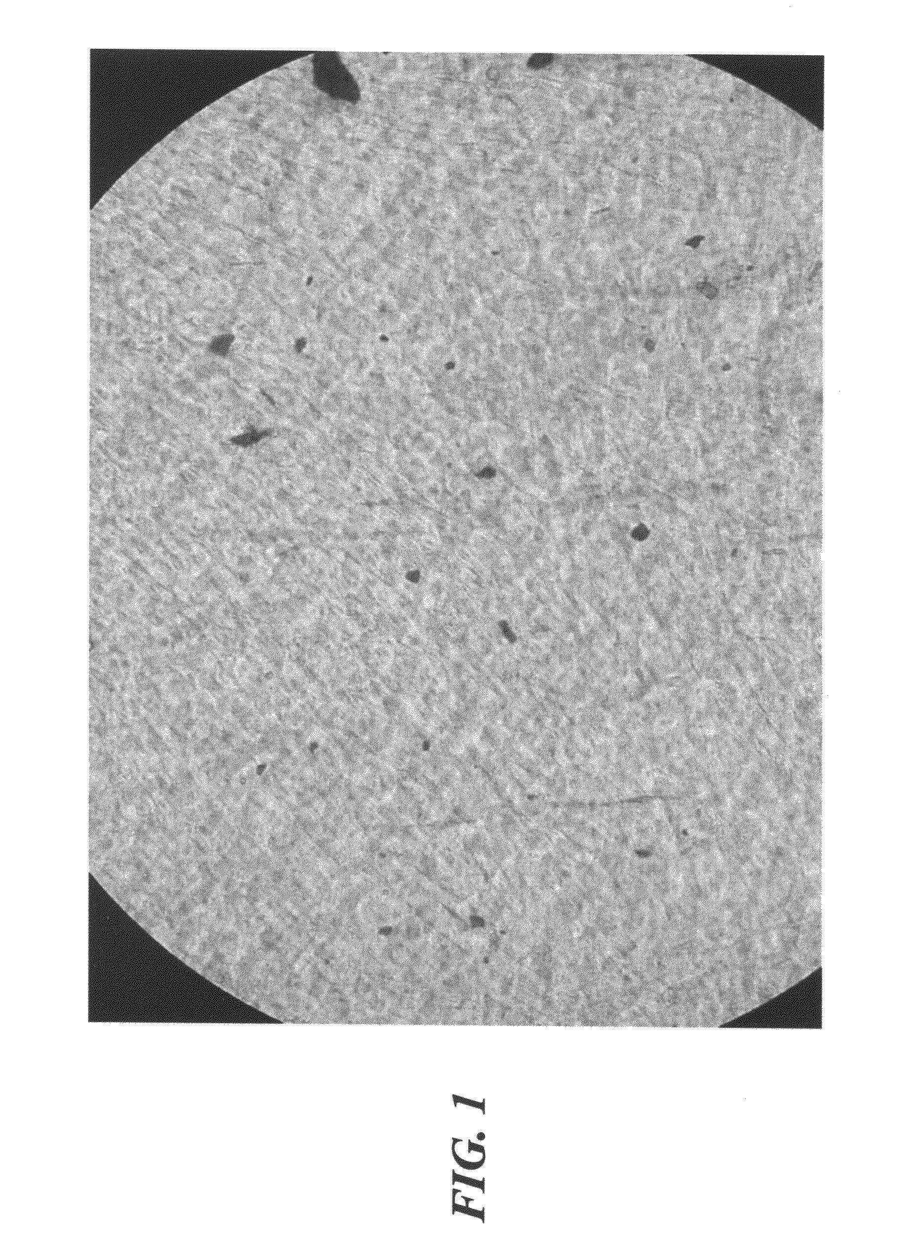
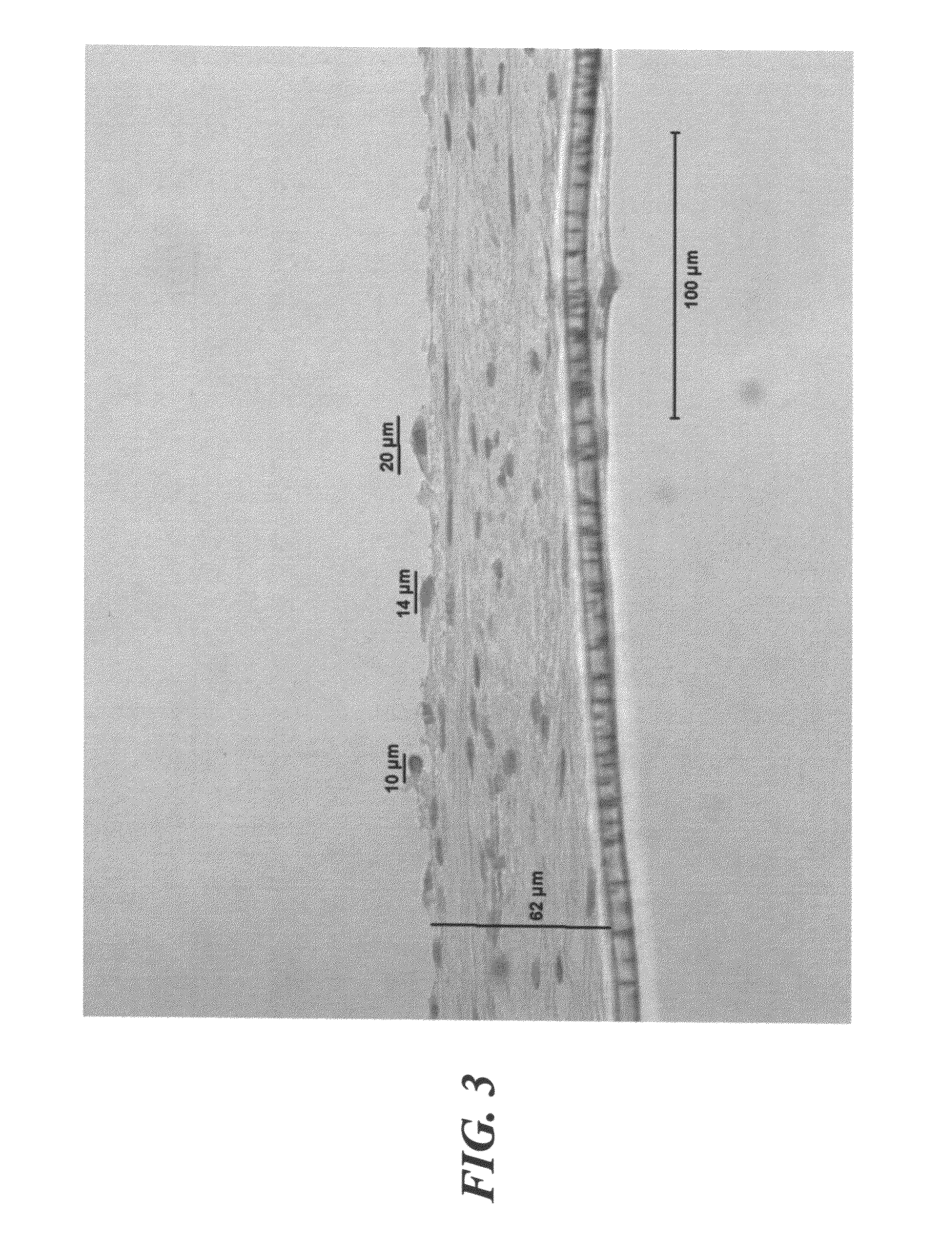
Production of Extracellular Matrix by Fibroblast Cells Isolated from the Dermis of Newborn Human Foreskin

[0154]Large numbers of fibroblast cells were isolated from the dermis of newborn human foreskin. Cells were proliferated in cell culture flasks fed with DMEM Medium with 10% Nu-Serum (or FBS or BCS) for several weeks. After a few passages, the fibroblast cells were centrifuged at 1000 RPMs, the supernatant decanted, and the cells pooled. These pooled fibroblast cells were resuspended in ALS Medium as described above in Example 1, then seeded into TRANSWELLS™ or BD FALCON™ / BIOCOAT™ Cell Culture Inserts or 6- or 12-well plates or other small containers suitable for in vitro cell culture, at superconfluent conditions, whereby cell density was between about 200,000 to greater than about 1,000,000 cells / cm2. The fibroblast cells were maintained at hyperconfluent conditions for about 3 weeks (or optionally about 1 week or longer than 10 weeks), during which time they were observed to p...


PUM

PropertyMeasurementUnit
tensile strengthaaaaaaaaaa
concentrationaaaaaaaaaa
thicknessaaaaaaaaaa

Abstract

The present invention is drawn to a 3-dimensional cell-produced scaffold construct comprising fibroblasts and the extracellular matrix that has been produced and arranged by these cells. The constructs promote the differentiation, dedifferentiation and / or transdifferentiation of cells and / or formation of tissue in vitro and in vivo, while at the same time promoting cell growth, proliferation, migration, and acquisition of in vivo-like morphology.

Description

CROSS REFERENCE TO RELATED APPLICATIONS[0001]This application is a U.S. regular application from U.S. Provisional Application Ser. No. 60 / 551,431 filed Mar. 9, 2004, the entire contents of which are hereby incorporated by reference herein.TECHNICAL FIELD[0002]The present invention relates to “living scaffolds” particularly “autogenic living scaffolds” (ALS), that comprise suitable living cells and the extracellular matrices these living cells produce. The invention also relates to the use of such autogenic living scaffolds as templates and supporting structures for growth of the same cells and tissue or the growth of different cells and different tissueBACKGROUND ART[0003]In the United States, millions of people are affected by tissue loss every year. Current treatments include tissue transfer from a healthy site in the same or another individual, use of medical devices to support the function of the lost tissue, or pharmacologic supplementation of the metabolic products of the lost...

Claims


Application Information

Patent Timeline
Patent Type & Authority Patents(United States)
IPC IPC(8): A01N63/00C12N5/00C12N5/071A61F2/10A61K45/00A61L27/38
CPCA61L27/3633C12N5/0658A61L27/3839A61L27/3886A61L27/3895C12N5/0671C12N5/0619A61L27/56A61L27/54A61L27/3834A61L27/3804A61L27/3687A61L27/3675A61L27/367A61L27/3654A61L27/3641A61L27/3683C12N2533/90A61L2430/32A61L2430/30A61L2430/28A61L2430/06A61L2430/00A61L2300/64C12N2513/00
Inventor AHLFORS, JAN-ERIC W.
Owner GENESIS TECH LTD
Why Eureka
Try Eureka