Transcoronary sinus pacing system, LV summit pacing, early mitral closure pacing, and methods therefor

a transcoronary sinus and pacing technology, applied in the field of transcoronary sinus pacing system, lv summit pacing, early mitral closure pacing, and methods therefor, can solve the problems of pacemaker syndrome, ventricular pacing, and very effective pacemaker pacing, so as to facilitate identification of position and orientation, reduce the time required for activation, and optimize the early closure of the mitral valve

Active Publication Date: 2011-11-29
GAUDIANI VINCENT
View PDF107 Cites 25 Cited by
  • Summary
  • Abstract
  • Description
  • Claims
  • Application Information

AI Technical Summary

Benefits of technology

[0009]The posterior summit of the left ventricle has been identified as the ideal location for an LV pacing lead because it optimizes early closure of the mitral valve in addition to reducing the time that it takes to activate the ventricle with BiV pacing. A permanent pacing electrode introduced by Seldinger technique that pierces the coronary sinus and achieves a stable location on the summit of the left ventricle is disclosed. A double lumen sheath that can place the pacing catheter in the coronary sinus and facilitate lodging it in the summit of the posterior LV is disclosed. A method for correlating angiographic coronary anatomy with live fluoroscopic pacing catheter placement is provided. Finally, a method for using intravascular ultrasound with pacing catheter placement is provided. These methods should help prevent damage to any coronary artery and prevent perforation of the epicardium (not shown) when the left ventricular summit (LV summit) is pierced.
[0012]In some configurations, the elongate sheath can be steerable by a guidewire. Additionally, the elongate sheath further provides radiopaque markers to facilitate locating the distal end of the device in situ. The elongate sheath can also have a teardrop cross-section which is used to determine orientation of the exit port relative to target anatomy. Furthermore, the pacing catheter is positioned anatomically in order to optimize early closure of the mitral valve. Positioning of the pacing catheter can reduce the time required to activate the left ventricle. As a result of these configurations, the pacing catheter is positioned in the heart irrespective of the anatomical position of the veins of the coronary sinus, while still optimizing performance. The pacing catheter can furthermore have a tip configured to pierce the coronary sinus.
[0014]Another aspect of the invention is directed to a left ventricular summit pacing device. The left ventricular summit pacing device comprises: an elongate sheath having a distal end, a proximal end and two lumen; and a pacing catheter the tip of which can perforate the coronary sinus and lodge in the LV summit. The elongate sheath is steerable by a guidewire, can provide radiopaque markers and / or have a teardrop cross-section. Additionally, the pacing catheter can be anatomically positioned in order to optimize early closure of the mitral valve. In some aspects, the pacing catheter is configured to reduce the time required to activate the left ventricle. The pacing catheter can be placed irrespective of the anatomical position of the veins of the coronary sinus and can have a tip configured to pierce the coronary sinus.
[0015]Still another aspect of the invention is directed to an early mitral valve closure pacing device. The early mitral valve closure pacing device comprises: an elongate sheath having a distal end, a proximal end and two lumen; and a pacing catheter having a pacing needle the tip of which can perforate the coronary sinus and lodge in the LV summit. The elongate sheath is steerable by a guidewire and provides one or more radiopaque markers to identify its orientation in the coronary sinus. The elongate sheath can also be configured to have a teardrop cross-section. The provision of radiopaque markers and cross-sectional shaping facilitates identification of the position and orientation of the device in situ. The pacing catheter can further be anatomically positioned in order to optimize early closure of the mitral valve and to reduce the time required to activate the left ventricle. The pacing catheter is configured for placement irrespective of the anatomical position of the veins of the coronary sinus. Additionally, the pacing catheter can be configured to provide a tip configured to pierce the coronary sinus.
[0016]An aspect of the invention is directed to a method for implanting a pacing device through the coronary sinus into the summit of the left ventricle. The method comprises the steps of: introducing a sheath; steering the sheath to a target location within the coronary sinus; lodging a distal end of the sheath beyond a posterior summit of the left ventricle; advancing a pacing lead through a lumen of the sheath to the target location within the coronary sinus; perforating the coronary sinus with the pacing lead; and removing the sheath. Additionally, the method can include the steps of advancing a right ventricular pacing catheter to a target location within the heart; and positioning an electrode at the target location within the heart. In some instance, an additional step of correlating an angiogram with live fluoroscopy is performed; or correlating an angiogram with live intravascular ultrasound. The pacing device is implanted through the coronary sinus into the summit of the left ventricle. Once implanted, the summit of the left ventricle can be activated to initiate closure of the mitral valve early in systole. Additionally, the cross-sectional shape of the device can be used in the method to facilitate steering and perforating.

Problems solved by technology

Ventricular pacing, however, while very effective in preventing deaths from such entities as complete heart block, has several problems associated with it.
One problem associated with ventricular pacing is pacemaker syndrome, which is a hemodynamic abnormality that can result when use of ventricular pacing is, for example, uncoupled from the atrial contraction.
It can also result from a less effective contraction caused by the abnormal mechanism of depolarization caused by the right ventricular pacing catheter.
In other patients, pacing induces a long term malfunction of the heart called cardiomyopathy that is caused by this slower depolarization of the heart and the associated decline in pump efficiency.
This asynchrony greatly reduces the efficiency of the heart in some patients with heart failure.
Additionally, one of the risks associated with placement of the device is the potential to damage the coronary sinus or coronary veins by, e.g., dissection or perforation.
Notwithstanding the clinical benefits of biventricular pacing, correctly placing the LV lead to achieve optimum performance may be difficult.
However, the surgical procedure requires a small incision in the chest and most cardiologists cannot do this and do not want to refer patients to surgeons.

Method used


Image

  • Transcoronary sinus pacing system, LV summit pacing, early mitral closure pacing, and methods therefor
  • Transcoronary sinus pacing system, LV summit pacing, early mitral closure pacing, and methods therefor
  • Transcoronary sinus pacing system, LV summit pacing, early mitral closure pacing, and methods therefor

Examples


Embodiment Construction

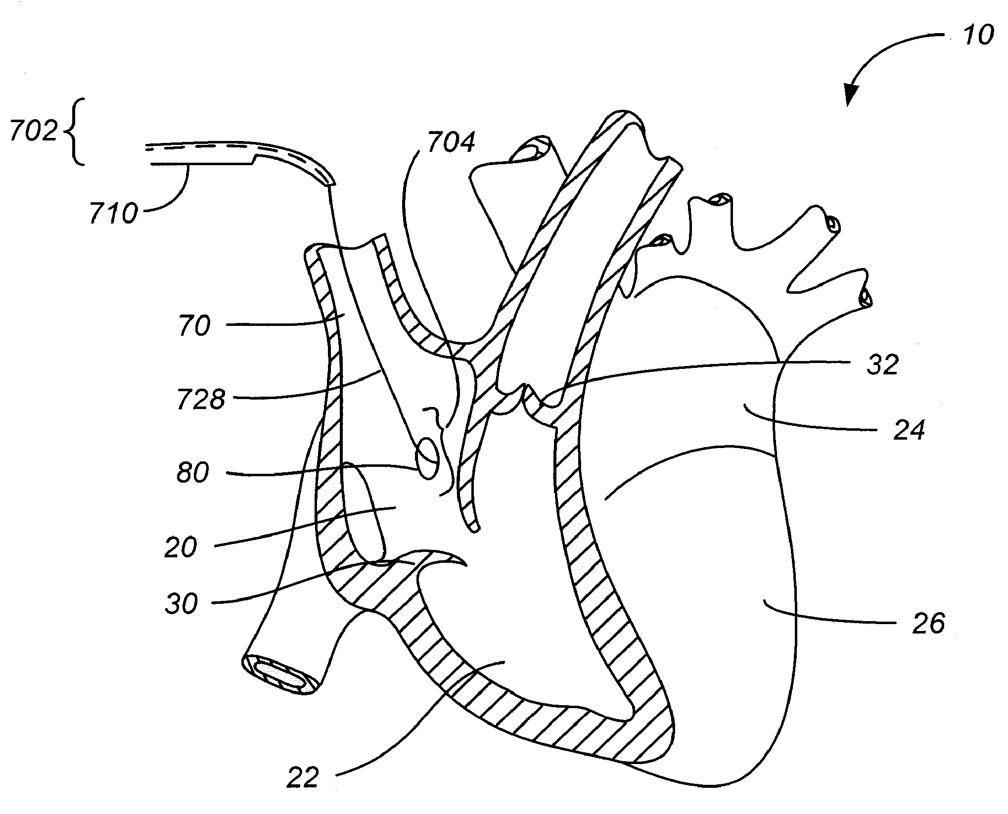
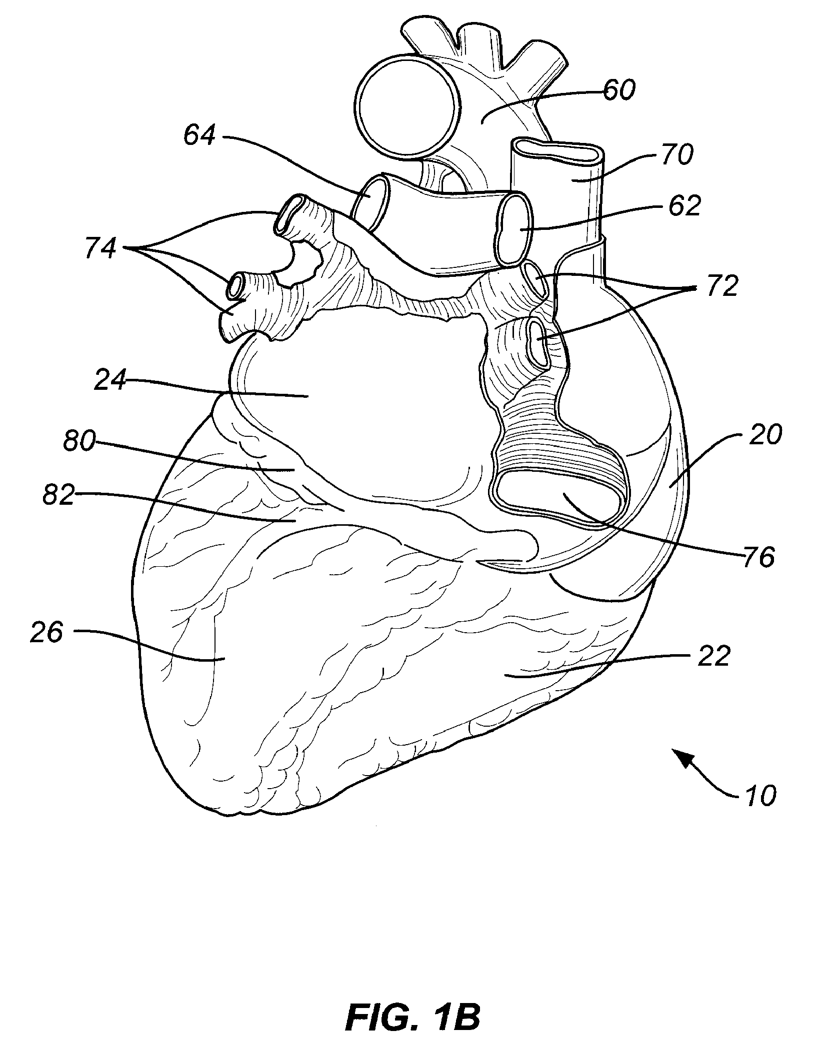
[0048]This system obviates the need for a separate lead placed in a coronary sinus vein 82 for transcoronary sinus pacing systems, as is currently practiced. The system relies on a the use of a single pacing catheter, which acts as an electrode lead, that is advanced through a guiding catheter to be positioned directly in, or near, the summit 28 of the left ventricle 26. Once positioned, the pacing catheter perforates the wall of the coronary sinus 66, locates the summit 28 of the left ventricle 26 a few millimeters from the place where the perforation occurs, and engages the summit 28 of the left ventricle 26 with an electrode tip. The actual location of perforation will be governed by patient specific anatomy and may vary slightly to avoid perforation of a coronary artery or ending up in the pericardium.

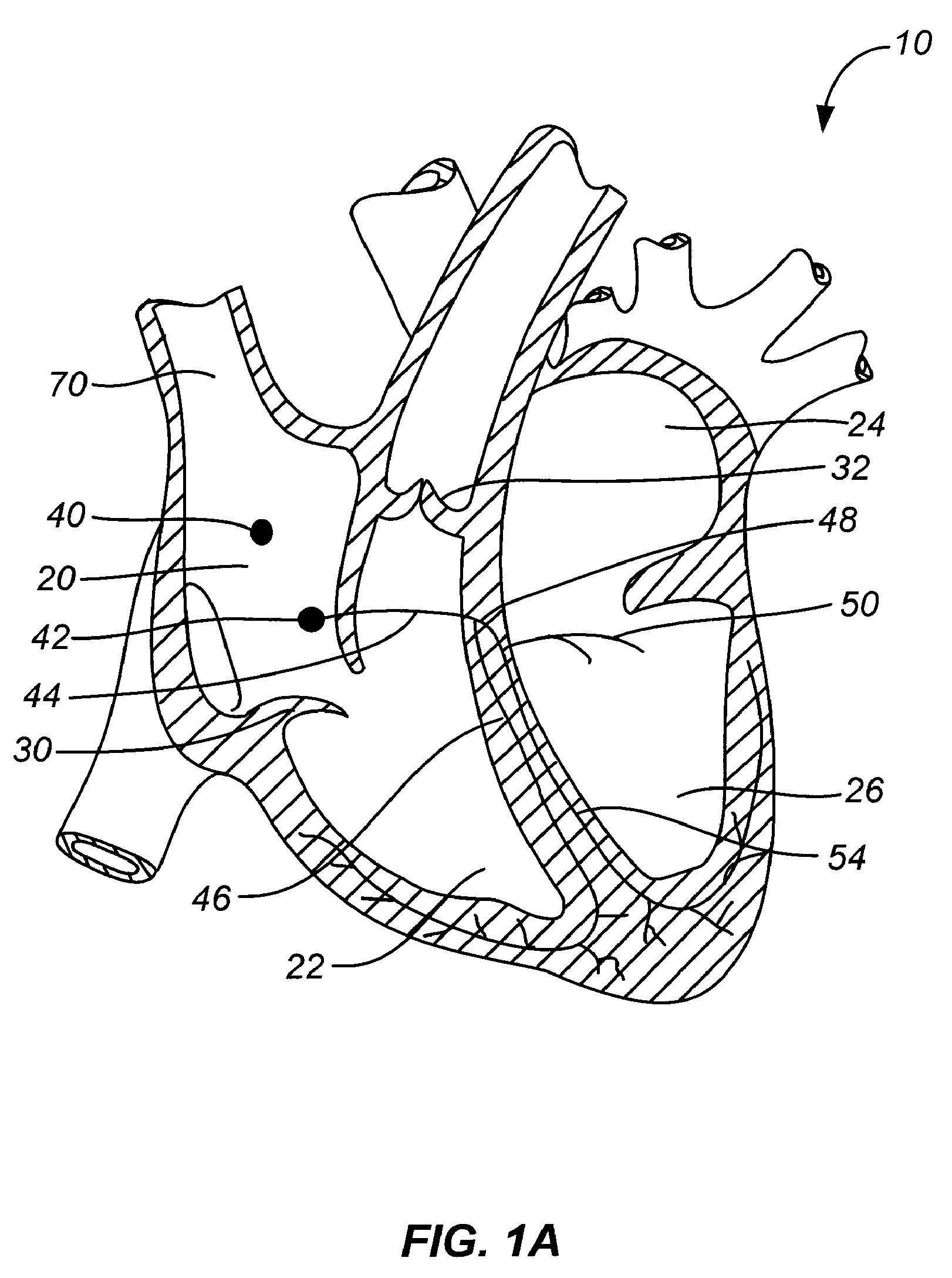
[0049]In order to appreciate the novelty of the invention, it is important to understand the basics of the human conduction system of the heart 10. The normal human conduction syst...


PUM


Abstract

A transcoronary sinus pacing system comprising a sheath having a lumen, a pacing catheter having a pacing needle, wherein the catheter can be advanced within the lumen and placed in the LV summit, and a right ventricular pacing device. A LV summit pacing device. An early mitral valve closure pacing device configured to operate with a right ventricular apex pacing device. A method for implanting a pacing device at a target coronary sinus tissue location, wherein the target can be the posterior LV summit. A method for achieving early closure of a mitral valve. A method for using visualization devices such as fluoroscopy or ultrasound and / or catheter features such as a radiopaque marker to locate a target location for LV pacing and to avoid piercing an artery or the pericardium when anchoring the LV pacing electrode.

Description

CROSS-REFERENCE[0001]This application claims the benefit of U.S. Provisional Application No. 60 / 828,708 filed Oct. 9, 2006, by Vincent A. Gaudiani entitled “Transcoronary Sinus Pacing System, LV Summit Pacing, Early Mitral Closure Pacing, and Methods Therefor,” and claims the benefit of U.S. Provisional Application No. 60 / 827,967, filed Oct. 3, 2006, by Vincent A. Gaudiani entitled “Transcoronary Sinus Pacing System, LV Summit Pacing, Early Mitral Closure Pacing, and Methods Therefor,” which are incorporated herein by reference in their entirety.BACKGROUND OF THE INVENTION[0002]Ventricular pacing has been a useful technique for at least 50 years, and transvenous pacing for nearly that long. In the transvenous pacing system the lead is placed from a vein, usually in the thorax, and threaded into the right ventricle 22 of the heart 10. The lead in the right ventricle 22 permits pacing and sensing within that chamber. Of course, pacing from the right ventricle 22 depolarizes the heart ...

Claims


Application Information

Patent Timeline
Patent Type & Authority Patents(United States)
IPC IPC(8): A61N1/05
CPCA61B17/3468A61M25/0082A61N1/0587A61N1/056A61B6/481A61B6/485A61B8/5292A61B19/5244A61B19/54A61B2019/5276A61M25/0021A61N1/3684A61N2001/0585A61M2025/0037A61B34/20A61B90/39A61B2090/378A61N1/36843
Inventor GAUDIANI, VINCENT A
Owner GAUDIANI VINCENT
Why Eureka
Try Eureka