Feature based neural network regression for feature suppression

a neural network and feature technology, applied in image enhancement, image analysis, instruments, etc., can solve the problems of difficult localization of other objects, difficult chest radiograph detection, and difficult detection of lung nodules, and achieve the effect of reducing the number of chest radiographs

Active Publication Date: 2012-06-19
RIVERAIN MEDICAL GROUP
View PDF3 Cites 13 Cited by
  • Summary
  • Abstract
  • Description
  • Claims
  • Application Information

AI Technical Summary

Problems solved by technology

The detection and localization, segmentation, of objects in images, both two dimensional and three dimensional, is commonly complicated by noise (both random and structured) and partial obscuration of objects of interest.
The detection of lung nodules, as well as other pathologies and objects of interest, whether based on computer aided detection (CAD) or human observers, in chest radiographs is challenging.
The detection and / or localization of other pathologies and objects of interest (e.g., catheters, feeding tubes, etc) in chest radiographs, particularly portable chest x-rays, is challenging and perhaps one the most difficult interpretation tasks in radiology.
Such difficulties may arise due to, for example, one or more of the following factors: poor patient positioning, imaging area and habitués, image latitude and dynamic range, poor x-ray penetration, and perhaps most significantly, the presence of obscuring bones.
The presence of the bone can lead to false diagnosis, false positives (FPs), false negatives and / or improper positioning of catheters and feeding tubes.
These difficulties may arise due to the projection of a three dimensional object onto a two dimensional image.
Even more significantly, overlapping bone may obscure the area underneath, resulting in a prominent source of false negatives.
Furthermore, the profile of the nodule and / or other relevant pathologies or structures (e.g., catheters), may be modified by the overlaying rib, which may result in more difficult interpretation tasks for both machines and practitioners.
The result can be noisy, and the example implementation only worked for bones away from the outer part of the lung field.
This means that the method may not perform well on data that does not look very similar to its training images.
Without feature extraction, a smooth approximation (good interpolation) is much harder to achieve.
Again, this implies that the approach may be susceptible to being too particular to its training images.
This is not to suggest that the technique will altogether fail, but only that it is more difficult to be confident in later predictions.
This approach would appear to have two major shortcomings: the first is that the prediction phase is entirely too computationally intensive and is likely inadequate.
The second is that the approach for image normalization is likely grossly inadequate.
Unfortunately, even at a coarse resolution, a few images can generate many pixels (large training set).
Therefore, for the routine to be even remotely practical, it would require a very sparse sampling of the training images.
However, sparse sampling of training images could lead to issues in prediction, as nearest neighbor methods are notoriously bad interpolators.
This would require a large value of K to compensate; however, too large a value of K leads to overly smoothed predictions (which would appear to be the case based on the images presented in the paper).
This step is potentially brittle in the presence of large non-anatomical artifacts (e.g., pacemakers) and allows for content outside the lung-field to have a heavy influence on pixel values inside.

Method used


Image

  • Feature based neural network regression for feature suppression
  • Feature based neural network regression for feature suppression
  • Feature based neural network regression for feature suppression

Examples


Embodiment Construction

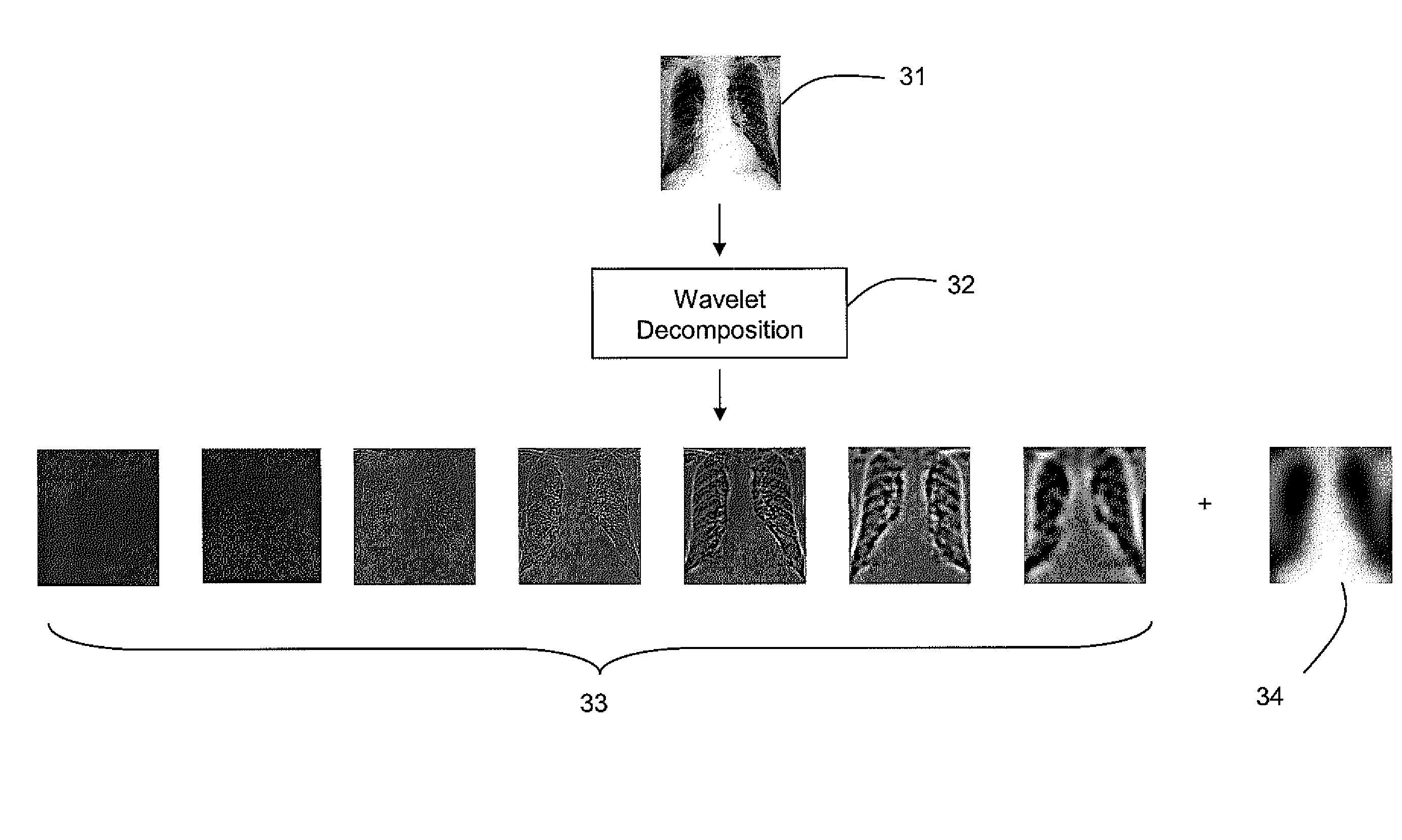
[0011]The developed bone suppression technique according to various embodiments of the invention may use dual energy (DE) data to generate a regression model for predicting the bone image. Along with a regression model that may be based on a set of robust, extracted features, the regression model may use a multi-layer perceptron (MLP) neural network architecture. Multiple feature representations may be used. Particular features may include, for example, in an exemplary embodiment of the invention:[0012]A 5 scale (redundant) wavelet decomposition using a third-order basic spline (B-spline) wavelet (this may be used to yield seven features, where five are from the details, one is from the low-pass residual, and the final is the original image);[0013]Five shape-index images, one for each of five scales, derived from harmonic second derivatives;[0014]Gaussian 4-jet at five separate scales.

[0015]To further explain the above concepts, the multi-scale representation provided by the wavelet...


PUM


Abstract

A method of obtaining one or more components from an image may include normalizing and pre-processing the image to obtain a processed image. Features may be extracted from the processed image. Neural-network-based regression may then be performed on the set of extracted features to predict the one or more components. These techniques may be applied, for example, to the problem of extracting and removing bone components from radiographic images, which may be thoracic (lung) images.

Description

CROSS-REFERENCE TO RELATED APPLICATION[0001]This application is a non-provisional patent application deriving priority from U.S. Provisional Patent Application No. 61 / 054,908, filed on May 21, 2008, and incorporated by reference herein.BACKGROUND[0002]The detection and localization, segmentation, of objects in images, both two dimensional and three dimensional, is commonly complicated by noise (both random and structured) and partial obscuration of objects of interest. The detection of lung nodules, as well as other pathologies and objects of interest, whether based on computer aided detection (CAD) or human observers, in chest radiographs is challenging. The detection and / or localization of other pathologies and objects of interest (e.g., catheters, feeding tubes, etc) in chest radiographs, particularly portable chest x-rays, is challenging and perhaps one the most difficult interpretation tasks in radiology. Such difficulties may arise due to, for example, one or more of the follo...

Claims


Application Information

Patent Timeline
Patent Type & Authority Patents(United States)
IPC IPC(8): G06K9/00
CPCG06T5/50G06T7/0012G06T7/0081G06T7/0087G06T2207/10116G06T2207/20064G06T2207/20084G06T2207/30008G06T2207/30064G06T7/11G06T7/143
Inventor KNAPP, JASONWORRELL, STEVE
Owner RIVERAIN MEDICAL GROUP
Why Eureka
Try Eureka