Imaging method for monitoring delivery of high dose rate brachytherapy

a brachytherapy and high dose rate technology, applied in the field of brachytherapy, can solve the problems of not providing direct monitoring in-situ of the cancer target, no accurate, reliable and reproducible method for determining the source,

Active Publication Date: 2012-10-23
JEFFERSON SCI ASSOCS LLC
View PDF6 Cites 8 Cited by
  • Summary
  • Abstract
  • Description
  • Claims
  • Application Information

AI Technical Summary

Benefits of technology

[0007]It is therefore an object of the present invention to provide an accurate, reliable and reproducible me

Problems solved by technology

The challenge in the use of this therapy is to verify that the highly radioactive 10 Ci pellet of Ir-192 is delivered accurately and reproducibly to one or multiple source dwell positions that are selected in the target tumor volume.
This method doe

Method used


Image

  • Imaging method for monitoring delivery of high dose rate brachytherapy
  • Imaging method for monitoring delivery of high dose rate brachytherapy
  • Imaging method for monitoring delivery of high dose rate brachytherapy

Examples


Embodiment Construction

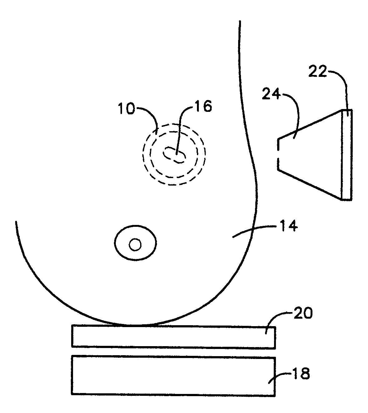
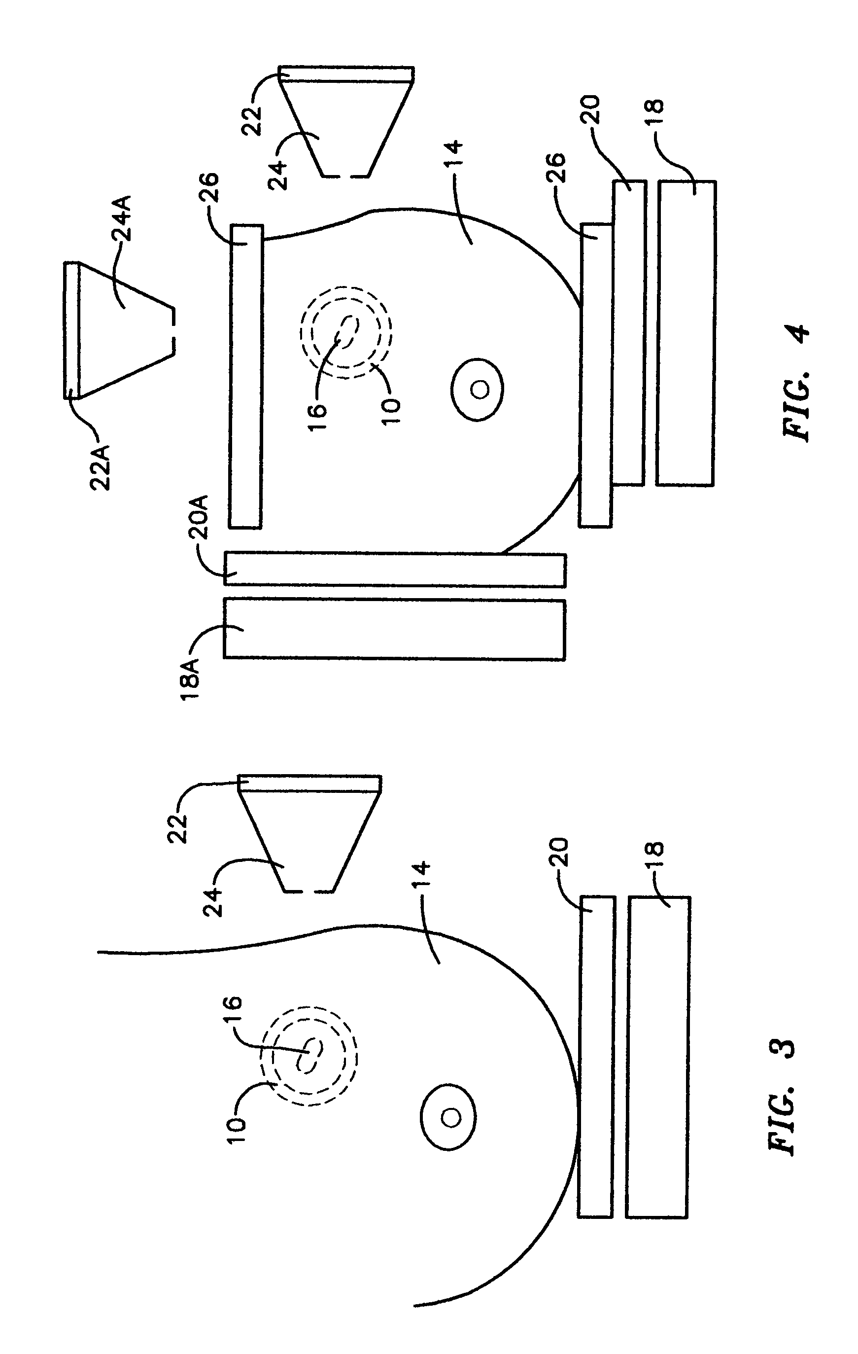
[0013]The novel method of the present invention makes either the breast tissue or the outline of the balloon visible to one or a set of gamma cameras implemented to image Tc-99m or similar low intensity radioactive pharmaceutical in order to see the location of the cavity / balloon while at the same time imaging the high intensity Ir-192 pellet source using a different or set of different gamma cameras built to image a high intensity radioactive source. By registering the relative positions of the post-surgical cavity / balloon and the Ir-192 pellet, the correct dose to the cavity tissue can be determined.

[0014]Referring now to the accompanying drawings that depict alternative embodiments of the method of the present invention, according to the embodiment depicted in FIG. 2, a first miniature gamma camera 18 including a parallel collimator 20 designed to image a low intensity pharmaceutical which is present in balloon 10 or breast tissue 14 after injection thereof in accordance with con...


PUM


Abstract

A method for in-situ monitoring both the balloon/cavity and the radioactive source in brachytherapy treatment utilizing using at least one pair of miniature gamma cameras to acquire separate images of: 1) the radioactive source as it is moved in the tumor volume during brachytherapy; and 2) a relatively low intensity radiation source produced by either an injected radiopharmaceutical rendering cancerous tissue visible or from a radioactive solution filling a balloon surgically implanted into the cavity formed by the surgical resection of a tumor.

Description

[0001]The United States of America may have certain rights to this invention under Management and Operating Contract DE-ACO5-060R23177 from the United States Department of Energy.FIELD OF THE INVENTION[0002]The present invention relates to brachytherapy and more particularly to a method for simultaneously imaging the radiation sources being applied and it relative location to tissue undergoing treatment.BACKGROUND OF THE INVENTION[0003]High dose rate brachytherapy has replaced low dose rate treatment of gynecological cancer and is being increasingly used for the treatment of breast, lung and prostate cancers. The challenge in the use of this therapy is to verify that the highly radioactive 10 Ci pellet of Ir-192 is delivered accurately and reproducibly to one or multiple source dwell positions that are selected in the target tumor volume. For multiple fractions delivered over 1 to 5 days / weeks, the reproducibility of source placement and the resultant dosimetry cannot, at this time,...

Claims


Application Information

Patent Timeline
IPC IPC(8): A61B5/05
CPCA61B6/12A61B6/4258A61B6/482A61N5/1015A61B6/022A61B6/5235A61N2005/1052
Inventor WEISENBERGER, ANDREW G.MAJEWSKI, STANISLAW
Owner JEFFERSON SCI ASSOCS LLC
Why Eureka
Try Eureka