
Chorionic villus sampling catheter

a catheter and chorionic villus technology, applied in the field of catheters, can solve the problems of deflection of the cannula tip from the desired location, rupture of the patient's membrane, and deflection of the cannula tip, and achieve the effect of convenient use and convenient use of ultrasound

Active Publication Date: 2012-12-04
WILLIAMS III JOHN +1
View PDF16 Cites 0 Cited by
  • Summary
  • Abstract
  • Description
  • Claims
  • Application Information

AI Technical Summary

Benefits of technology

"This patent is about an improved catheter for sampling areas of interest in the uterus, such as for chorionic villus sampling or other medical procedures. The catheter is made of a flexible plastic cannula and a flexible stainless steel obturator with a molded handle. It can be easily guided using ultrasound and can be inserted through the cervix into the uterus during pregnancy. The catheter can be used with a visualization device or method to aid in guidance. After placement, a sample of the area of interest can be removed by applying pressure to the catheter and withdrawing it from the uterine cavity. The technical effects of this invention include improved accuracy and efficiency in sampling areas of interest in the uterus, as well as improved safety and ease of use for the operator."

Problems solved by technology

The Martin patent mentions that a drawback of this catheter is the aluminum obturator tends to flex the curved portion of the cannula when removed, thus deflecting the cannula tip from the desired location.
Likewise, with this catheter the stiffness of the obturator causes deflection of the cannula tip during withdrawal of the obturator.
Additionally, the stiffness of the catheter would often cause ruptures in patients' membranes.
Once again, when the obturator is removed its relative stiffness will cause flexing in the cannula and movement of the tip.
Therefore, it has been previously disclosed that biopsy catheters for use in the CVS procedure, which use an obturator having greater stiffness than the cannula, may suffer from the disadvantage of tip displacement when the obturator is withdrawn.
However, this catheter was found not to work in all cases because the preset curvature angle did not fit the anatomy of some patients.
The stiffness of this catheter, however, was found by some practitioners to be undesirable.
Because the handle is flat, it is difficult to manipulate between the fingers of the physician / operator when inserting the catheter to the desired location to collect the sample.
The difficulty in manipulating this flat handle has been found to make placement of the catheter less precise in certain anatomical positions of the pregnant uterus.

Method used


Image

  • Chorionic villus sampling catheter
  • Chorionic villus sampling catheter
  • Chorionic villus sampling catheter

Examples


Embodiment Construction

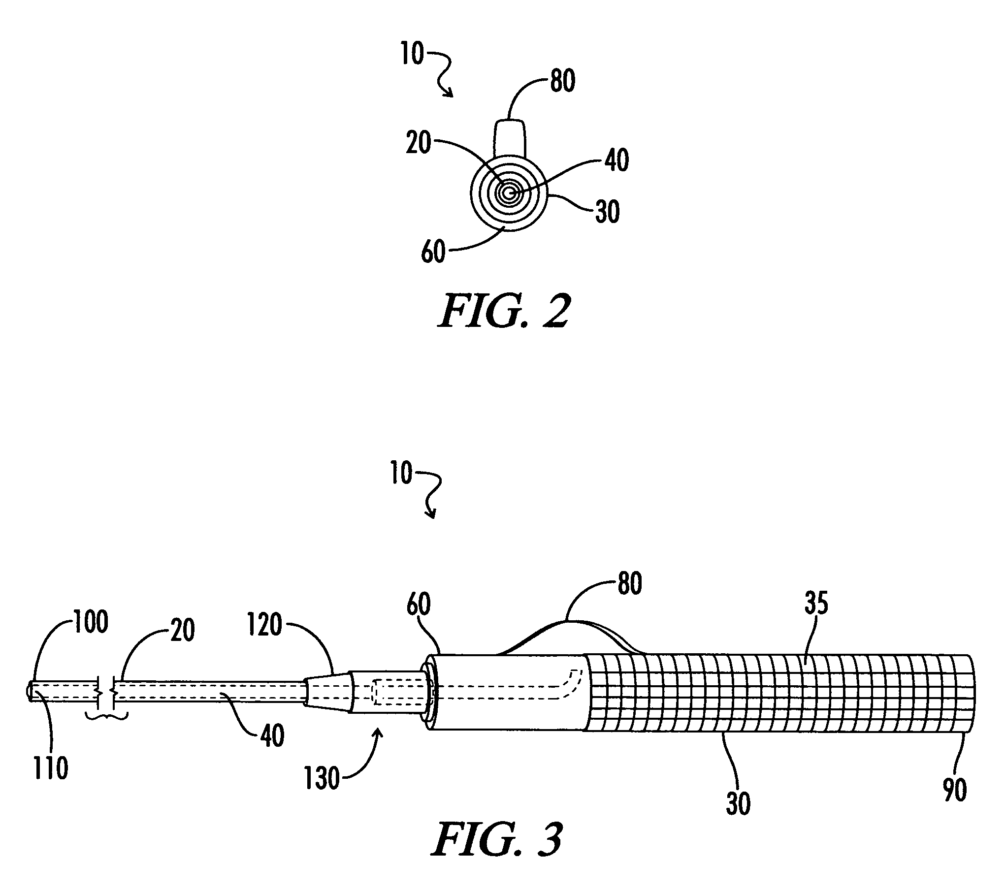
[0014]The present invention is directed to a catheter for use in sampling an area of interest, such as the chorionic villus sampling procedure or other uterine biopsy medical procedures, comprising a cannula readily adjustable by the operator to conform to the patient's anatomy and that remains in place when the obturator is removed to collect the sample, and an obturator with a generally cylindrical handle and steering guide to allow the operator ease of manipulation and placement of the catheter at the area of interest, for example, the chorion frondosum.

[0015]By way of explanation and example, the present invention will be described in detail below. It is to be understood, however, that the present invention is not limited to the specific structure described herein as will be evident to one of ordinary skill in the relevant art.

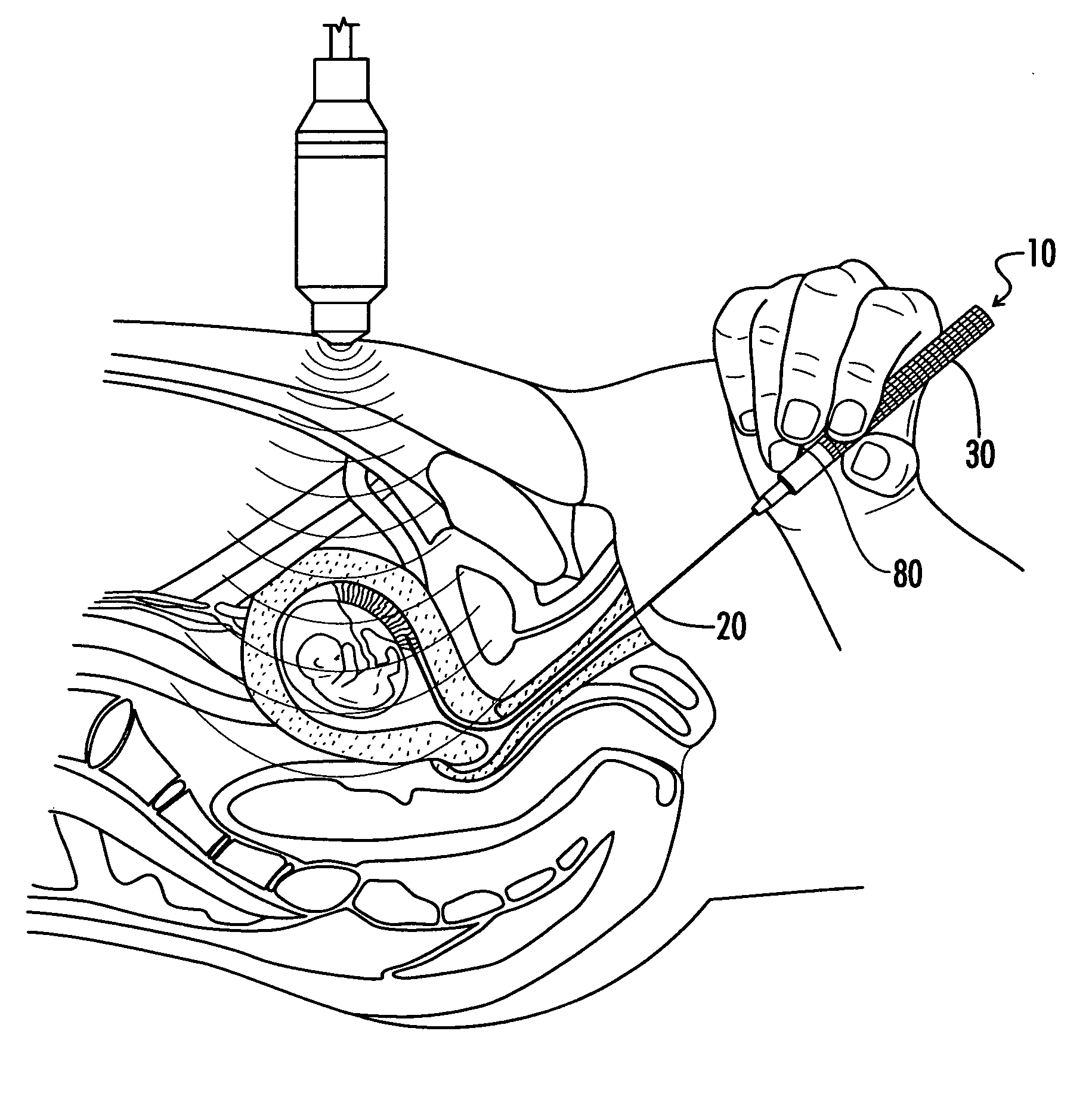
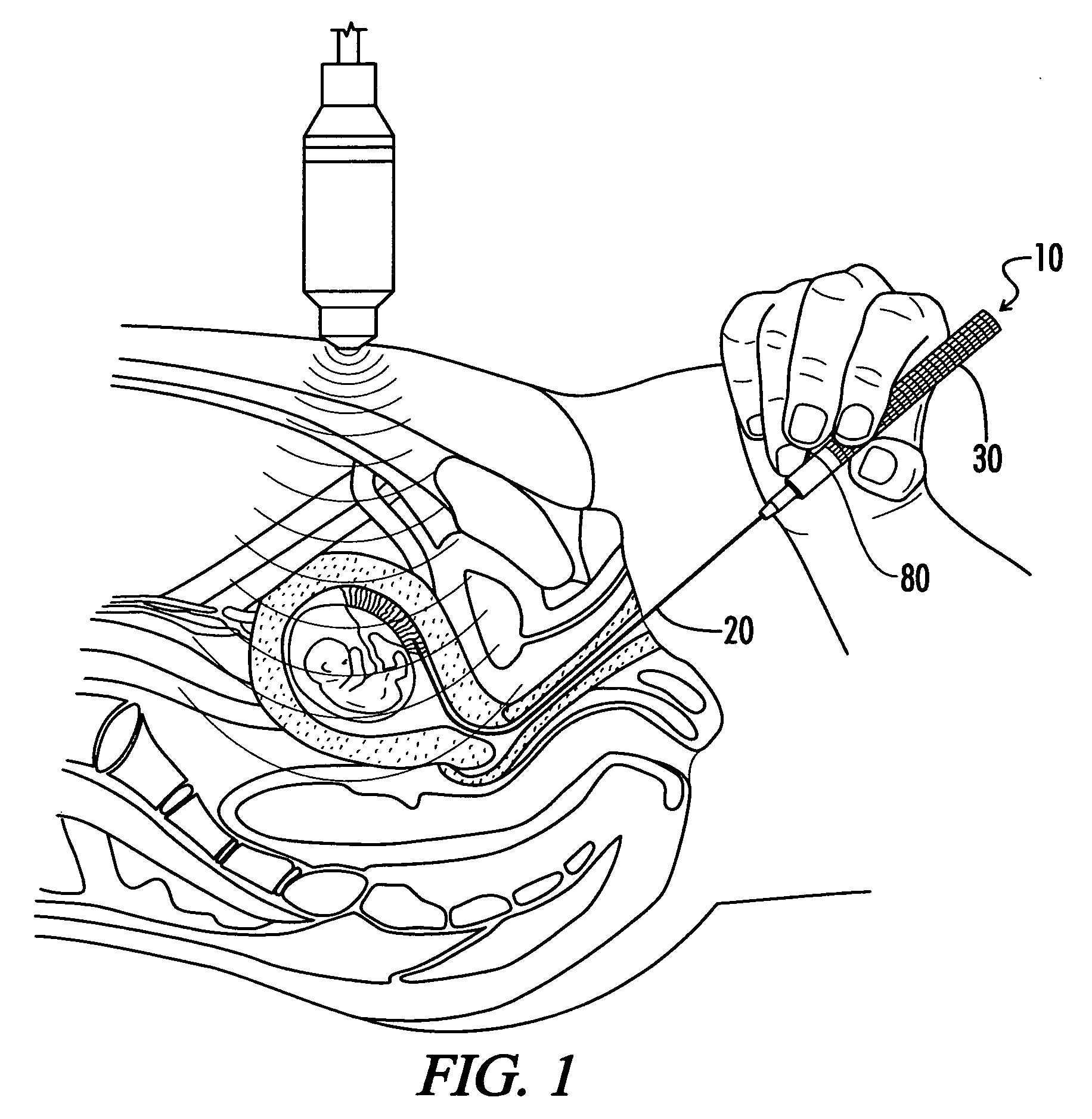
[0016]FIG. 1 is a diagrammatic view of an embodiment of the catheter of the present invention being inserted with the aid of a visualization or imaging de...


PUM


Abstract

A catheter with a guide wire and handle is provided for sampling an area of interest, such as chorionic villi. The catheter contains a cannula of flexible plastic that is non-pyrogenic and latex-free, and a flexible obturator with an elongate cylindrical handle. The cannula is echogenic and can be easily visualized with visualization or imaging device. In an embodiment, the catheter may be curved manually by the operator to conform to the patient's anatomy and it is inserted through the cervix into a pregnant uterus at 10-13 weeks gestation and placed into the developing placenta under ultrasound guidance and a guide mechanism provided on the handle. In this embodiment, once the catheter has been placed, the obturator is withdrawn and a sample of chorionic villi is removed by applying appropriate pressure to the proximal end of the cannula and slowly withdrawing the cannula from the uterine cavity.

Description

[0001]Related U.S. Application: Provisional application No. 61 / 131,201, filed on Jun. 6, 2008.FIELD OF INVENTION[0002]The present invention is directed to catheters for use in the chorionic villus sampling medical procedure.BACKGROUND OF THE INVENTION[0003]Chorionic villus sampling (CVS) is an established method for prenatal diagnosis of genetic diseases and birth defects in the first trimester of pregnancy, which has been in use since the 1980s. The procedure entails retrieving a sample of the chorionic villus or placental tissue and testing it. CVS is an alternative to genetic amniocentesis, a procedure used since the 1960s for essentially the same purpose, and may be performed sooner. Amniocentesis typically involves inserting a thin needle through the patient's abdominal wall through the wall of the uterus into the amniotic sac under ultrasound guidance during the 16-20 week of pregnancy. The CVS procedure may be carried out 10-13 weeks after the patient's last menstrual period....

Claims


Application Information

Patent Timeline
Patent Type & Authority Patents(United States)
IPC IPC(8): A61B10/00
CPCA61B10/0291A61B17/4241A61B2017/3413A61B2017/3456
Inventor WILLIAMS, III, JOHNWILLIAMS, JENNIFER MARIE
Owner WILLIAMS III JOHN
Learn More