Human trophoblast stem cells and use thereof

a technology of trophoblast stem cells and stem cells, which is applied in the field of isolated preparation of human trophoblast stem cells, can solve the problems of limited trophectoderm, inability to know the nature of embryo-derived signals, and failure to routinely culture mouse trophoblast stem cells for long-term cultur

Active Publication Date: 2006-09-21
ACCELERATED BIOSCI
View PDF2 Cites 29 Cited by
  • Summary
  • Abstract
  • Description
  • Claims
  • Application Information

AI Technical Summary

Benefits of technology

[0022] This invention further provides a composition for use in the treatment of patients suffering from habitual abortion and/or IVF failure comprising monocloned antibodies, antagonists and other inhibitors to regulate bFGF, Oct4, fgfr-2, and FGF4 levels. Th

Problems solved by technology

However, the trophectoderm is exclusively restricted to form the fetal portion of the placenta and the trophoblast giant cells.
However, the nature of the embryo-derived signal was not known and all attempts at routine long-term culture of mouse trophoblast stem cells have been unsuccessful (U.S. Pat. No. 6,330,349).
This may result from defects in the ICM and its endoderm derivatives.
Use of feeder cells for culturing the human embryonic stem cell (hESC) lines produces mixed cell population that require

Method used


Image

  • Human trophoblast stem cells and use thereof
  • Human trophoblast stem cells and use thereof
  • Human trophoblast stem cells and use thereof

Examples


example 1

Primary Culture and Isolation of hTS Cells from Trophoblastic Villi

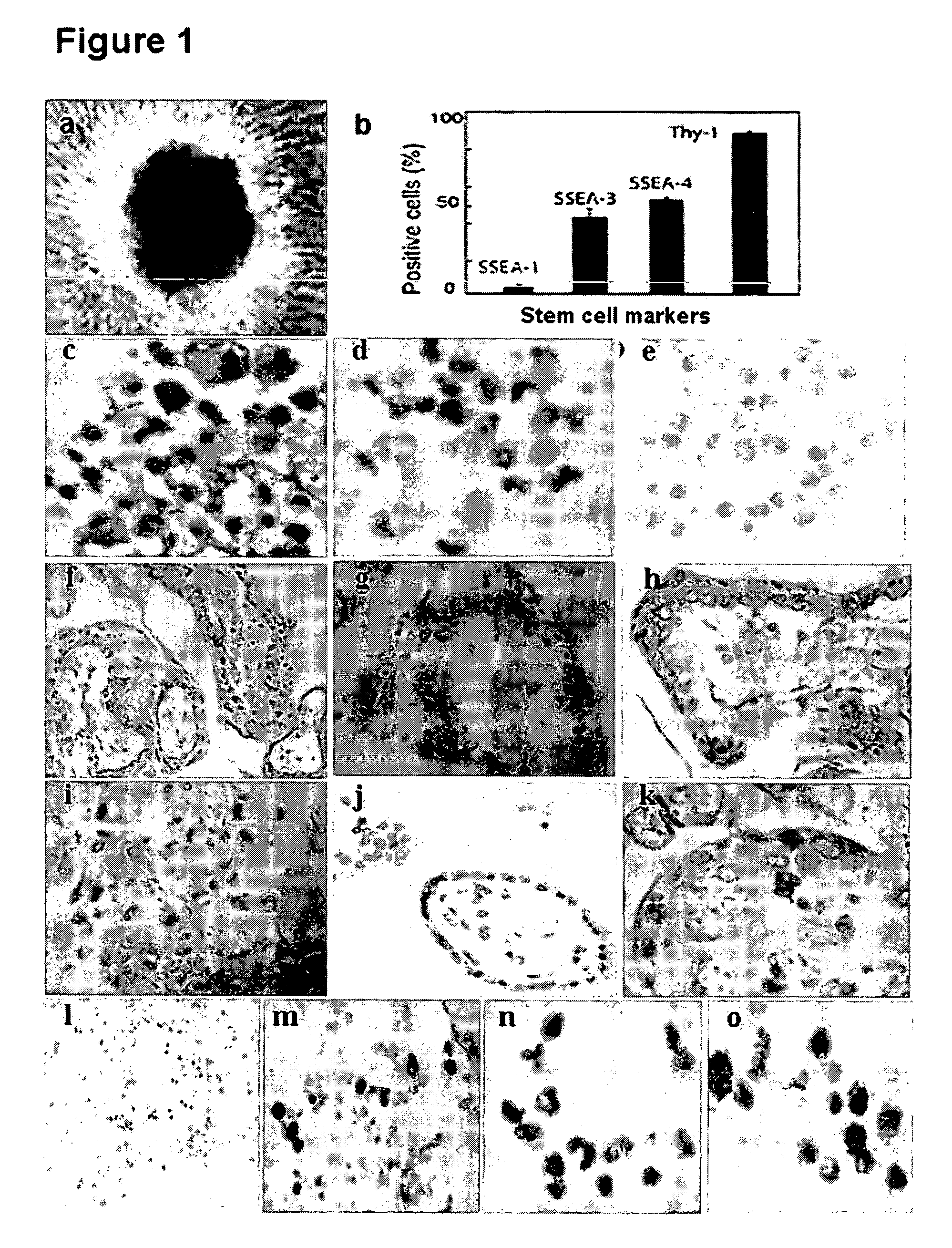
[0065] Using a laparoscopy, eight eligible samples (4-5 weeks post-fertilization) were obtained from the gynecologic unit (FIG. 10 a). The samples were used for cell culture and for immunohistochemical study. Fresh villi (FIG. 10 b) were mechanically dissected, washed, and cultured with medium in the absence of bFGF. Prior to differentiation, only few EBs appeared on day one of the culture (FIG. 1 a). After a week, EBs count increased to 30-40 in number with identical morphological features described in previous studies (M. Amit et al., Dev. Biol., 227, 271, (2000); M. J. Shambloott et al., Proc. Natl. Acad. Sci. USA., 98, 113, (2001)).

[0066] Early trophoblastic villi were obtained from the unruptured tubal ectopic pregnant mass (gestational age: 6-7 weeks) (FIG. 10 a) under laparoscopic surgery. Immediately, villous tissues were washed by normal saline (37° C.) to get rid of blood and dissected in serum free α-ME...

example 2

Immunocytochemical Study of Differentiated hTS Cells

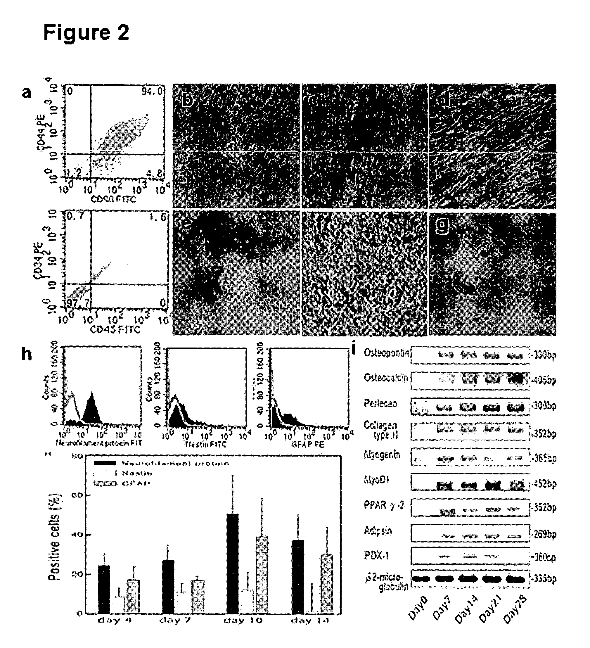
[0070] At passage 6, cells were cultured as a monolayer in basal medium (α-MEM containing 20% FBS and 10 ng / ml bFGF) in 3.5 cm culture dishes. After cells grew to 70% in confluence, culture medium was changed for variable differentiations. The evaluations were made at day 7, 14, 21, and 28 after drug induction. For osteogenic differentiation, cells were grown in conditioned medium (Table 1).

TABLE 1Recipes used for specific cell phenotype differentiations of hTS cellsDifferentiationsReagentsOsteogenesisα-MEM containing 20% FBS, 10 μg / ml bFGF,0.1 μM dexamethasone, 10 mMβ-glycerolphosphate, and0.2 mM ascorbic acidChondrogenesisα-MEM containing 10% FBS, 1% antibiotic / antimycotic,6.25 μg / ml insulin, 10 ng / ml TGF-β1, and50 nM ascorbate-2-phosphateMyogenesisα-MEM containing 10% FBS, 10 μg / ml bFGF,0.1 mM dexamethasone, 50 mM hydrocortisone, and 5%horse serumAdipogenesisα-MEM containing 20% FBS, 10 μg / ml bFGF,10 μg / ml insulin, 1 μM dexa...

example 3

RT-PCR

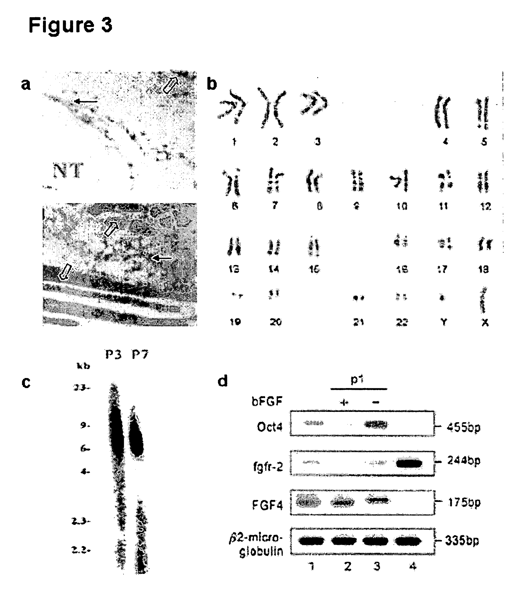
[0080] Extraction of total RNA from the hTS cells (105 to 106) at various passages of culture and term placental tissues was carried out using the TRIzol kit according to the manufacturer's instructions (Invitrogen, Carlsbad, Calif.). Reverse transcription was performed using 1 μg. RT reactions were performed using Ready-to-Go, RT-PCR Beads kit (Amersham Biosciences, Buckinghamshire, UK). One μg of total RNA was reverse transcribed to cDNA. The cDNA product, corresponding to 0.2 μg of total RNA, was used for PCR amplification. The primers used were shown in Table 2.

TABLE 2Primer sequences used for RT-PCROsteopontin(5′- CTAGGCATCACCTGTGCCATACC -3′forward and5′- CAGTGACCAGTTCATCAGATTCATC -3′reverse)Osteocalcin(5′- CGCAGCCACCGAGACACCAT -3′forward and5′- GGGCAAGGGCAAGGGGAAGA -3′reverse)Perlecan (PRLN)(5′- CATAGAGACCGTCACAGCAAG -3′forward and5′- ATGAACACCACACTGACAACC -3′reverse)Collagen type┌┐(5′- ACGGCGAGAAGGGAGAAGTTG -3′forward and5′- GGGGGTCCAGGGTTGCCATTG -3′reverse)Myogenin...


PUM


Abstract

Existence of human trophoblast stem (hTS) cells has been suspected but unproved. The isolation of hTS cells is reported in the early stage of chorionic villi by expressions of FGF4, fgfr-2, Oct4, Thy-1, and stage-specific embryonic antigens distributed in different compartments of the cell. hTS cells are able to derive into specific cell phenotypes of the three primitive embryonic layers, produce chimeric reactions in mice, and retain a normal karyotype and telomere length. In hTS cells, Oct4 and fgfr-2 expressions can be knockdown by bFGF. These facts suggest that differentiation of the hTS cells play an important role in implantation and placentation. hTS cells could be apply to human cell differentiation and for gene and cell-based therapies.

Description

RELATED APPLICATIONS [0001] This application claims priority from U.S. Provisional Patent Application Ser. No. 60 / 655,747 which was filed on Feb. 24, 2005.BACKGROUND OF THE INVENTION [0002] 1. Field of the Invention [0003] This invention relates to an isolated preparation of human trophoblast stem cells and use thereof. [0004] 2. Description of the Related Art [0005] In mammals, the earliest developmental decision specifies the trophoblast cell lineage. In mice, this lineage appears at the blastocyst stage as the trophectoderm, a sphere of epithelial cells surrounding the inner cell mass (ICM) and the blastoceol. After implantation, the ICM gives rise to the embryo proper and some extraembryonic membranes. However, the trophectoderm is exclusively restricted to form the fetal portion of the placenta and the trophoblast giant cells. The polar trophectoderm (the subset of trophectoderm in direct contact with the ICM) maintains a proliferative capacity and gives rise to the extraembryo...

Claims


Application Information

Patent Timeline
IPC IPC(8): C12N5/08A61K35/12C12N5/071C12N5/073C12N5/0735
CPCA61K35/12C12N5/0605C12N5/0606C12N5/0677C12N2500/34C12N2500/38C12N2500/44C12N2501/115C12N2506/02C12N2533/54G01N33/5073A61K35/39A61K35/54A61K39/395
Inventor LEE, JAU-NANLEE, TONYLEE, YUTA
Owner ACCELERATED BIOSCI
Why Eureka
Try Eureka