
Multimodal detection of tissue abnormalities based on raman and background fluorescence spectroscopy

a tissue abnormality and background fluorescence spectroscopy technology, applied in the direction of dianostics using fluorescence emission, medical automation diagnosis, sensors, etc., can solve the problems of systemic metastasis and death, invasive and impractical screening of high-risk patients, and more extensive and disfiguring surgery becomes necessary

Active Publication Date: 2012-12-04
BRITISH COLUMBIA CANCER AGENCY BRANCH
View PDF36 Cites 20 Cited by
  • Summary
  • Abstract
  • Description
  • Claims
  • Application Information

AI Technical Summary

Problems solved by technology

However, if detected late, more extensive and disfiguring surgery becomes necessary.
If treatment for malignant melanoma is commenced too late, systemic metastasis and death can occur.
Excisional biopsy currently remains the most reliable diagnostic approach for the early detection of skin cancer, but is invasive and impractical for screening high-risk patients who may have multiple suspicious lesions.
Many unnecessary biopsies are done, at considerably cost to the health care system.
Moreover, some needed biopsies may not be performed because of a failure to recognize a cancer.
If a tumor has margins that are poorly defined, it may be necessary to perform repeated biopsy procedures from multiple sites in a time-consuming, expensive, and tedious procedure known as Mohs micrographic surgery.
This approach has improved the registration, recording, and documentation of skin lesions, but has not yet significantly improved the accuracy of non-invasive diagnoses.
FT-Raman systems take up to ½ hour to acquire a spectrum and are bulky and not portable, and therefore are of limited clinical utility.
A problem with the evaluation of pigmented lesions, including melanoma and its precursors, by reflectance or visible fluorescence techniques is that melanin is a strong light absorber throughout the ultraviolet and visible spectrum.
This results in weak spectra and “black hole” images that provide little clinically useful information.
To date, none of the diagnostic methods described in the publications listed above have been widely adopted for use in tissue screening.

Method used


Image

  • Multimodal detection of tissue abnormalities based on raman and background fluorescence spectroscopy
  • Multimodal detection of tissue abnormalities based on raman and background fluorescence spectroscopy
  • Multimodal detection of tissue abnormalities based on raman and background fluorescence spectroscopy

Examples


application example # 2

APPLICATION EXAMPLE #2

[0121]As shown in FIG. 11, some features of Raman spectra are different between normal to benign (compound nevus) and malignant (melanoma) skin diseases. Curve 104 is a Raman spectrum of the volar forearm normal skin of a subject of African descent. Curve 105 is a Raman spectrum of a benign compound pigmented nevus. Curve 106 is a Raman spectrum of a malignant melanoma. Curve 107 is a Raman spectrum of normal skin adjacent the melanoma of curve 106. One can see significant differences between these curves. The 1445 cm−1 peak is not visible in the malignant melanoma spectrum 106 but can be seen in both the normal black skin spectrum 104 and the benign compound nevus spectrum 105. The 1269 cm−1 peak is present in the malignant melanoma spectrum 106 and in the normal black skin spectrum 104 but not in the benign compound nevus spectrum 105. Features of these curves may be used together with features of NIR autofluorescence which forms a background to these curves ...

application example # 3

APPLICATION EXAMPLE #3

[0122]Some methods of the invention provide a plurality of classification functions. Such methods may involve selecting the one of the classification functions most appropriate for classifying the tissue section involved. For example, the classification functions may include classification functions for any one or more of:[0123]a number of different pathologies (such as, for example, two or more of basal cell carcinoma (BCC), squamous cell carcinoma (SCC), melanoma, actinic keratosis, seborrheic keratosis, sebaceous hyperplasia, keratoacanthoma, lentigo, melanocytic nevi, dysplastic nevi, and blue nevi);[0124]a number of different tissue types (such as, for example, two or more of skin, lung tissue, other epithelial tissues, such as the bronchial tree, the ears nose and throat, the gastrointestinal tract, the cervix, and the like);[0125]a number of different skin types (for example, one classification function may be provided for use with subjects having lightl...

application example # 4

APPLICATION EXAMPLE #4

[0130]A patient has a condition such as dysplastic nevus. The condition causes many nevi at various locations on the patient's body. The patient visits his physician who needs to decide whether it is necessary to take a biopsy of any of the nevi and, if so, which ones. There are enough nevi that it is not desirable or practical to take biopsies of all of the nevi.

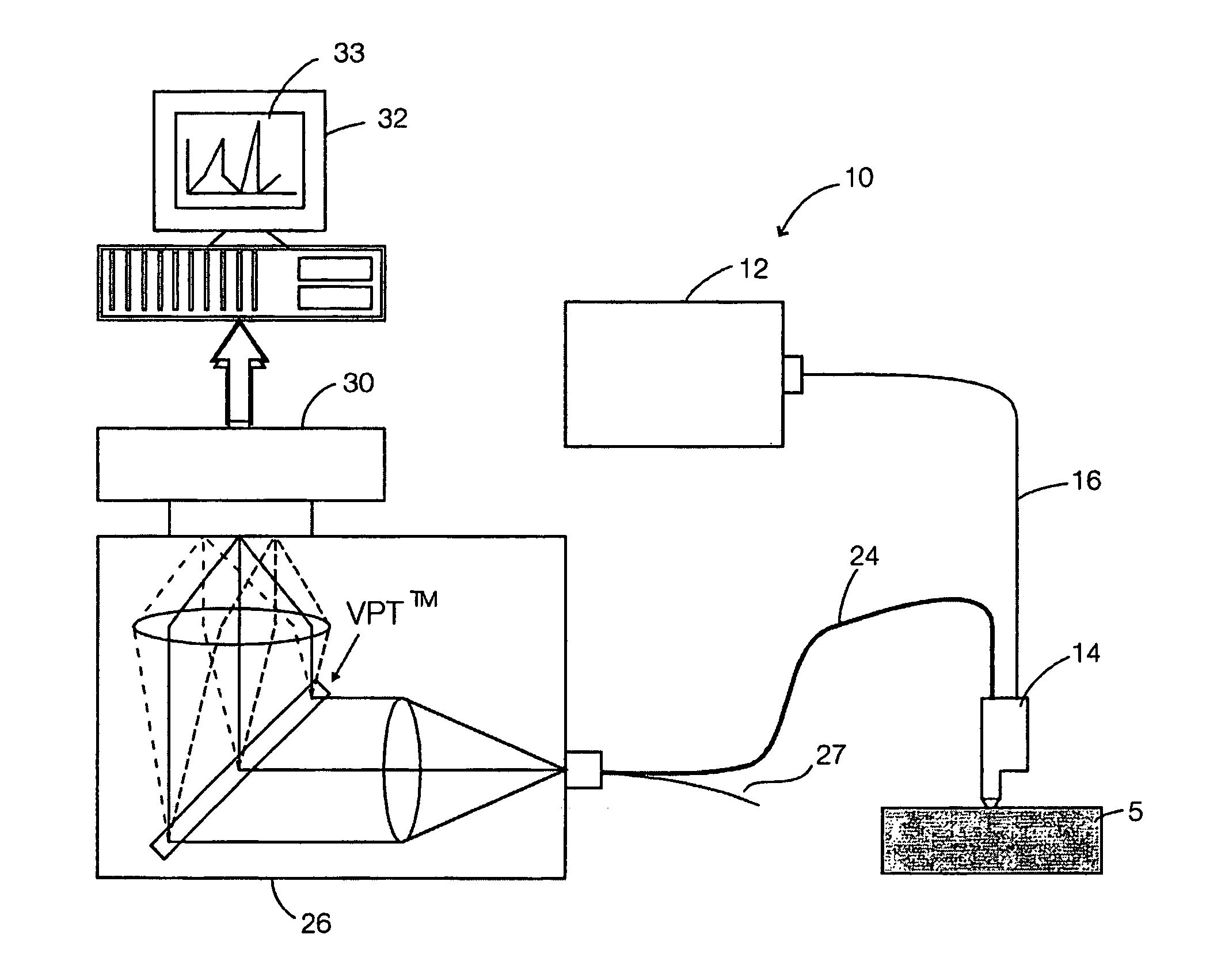
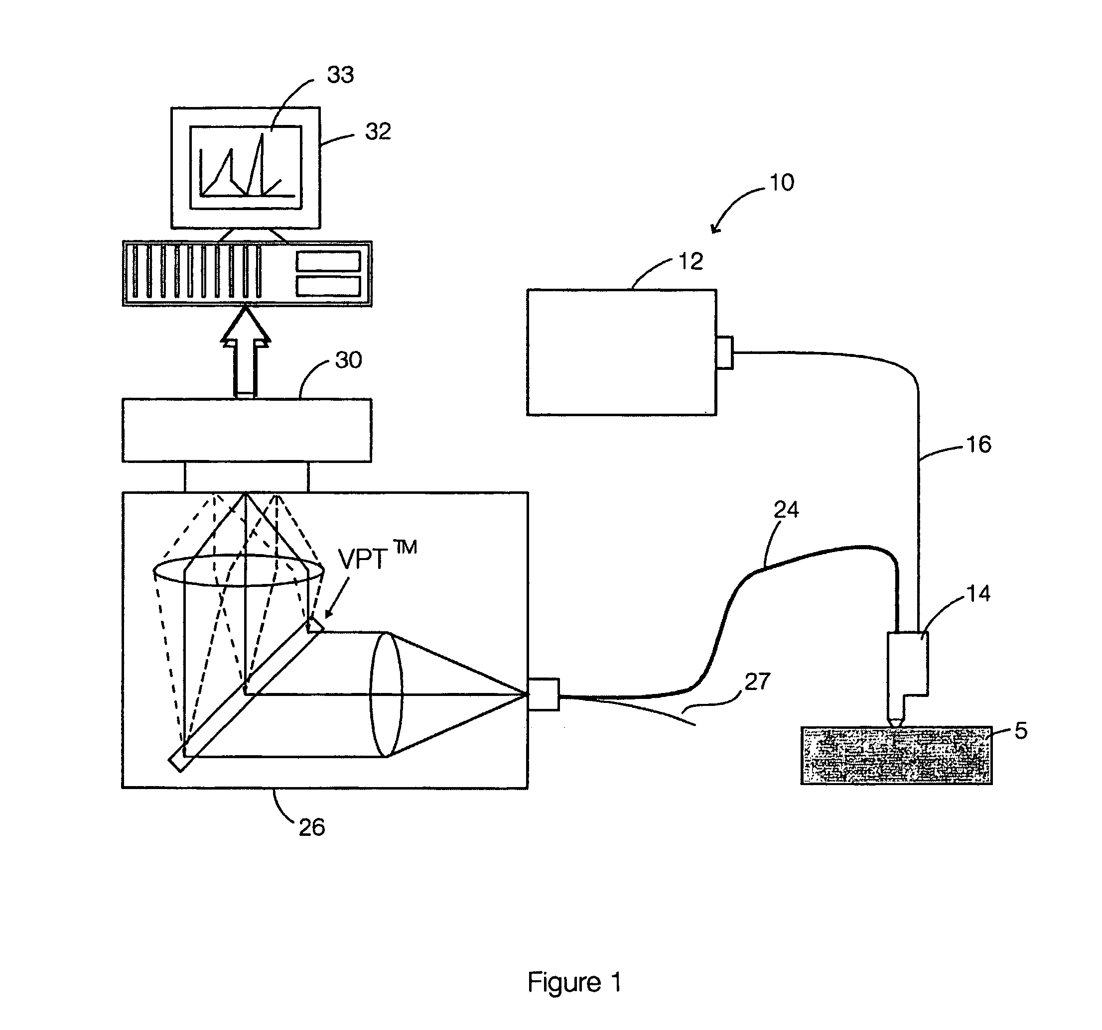
[0131]The physician obtains a NIR spectrum for each of the nevi to be investigated. The NIR spectra include both Raman features and NIR fluorescence features. The spectra may be obtained, for example, with apparatus as described above and shown in FIG. 1. The physician can place the probe of the apparatus on each nevus in turn and then trigger the acquisition of a spectrum by activating a control. For example, the physician may press a button when the probe is over a nevus and then hold the probe over the nevus until a spectrum has been acquired. The apparatus may generate a signal, such as an audible ...


PUM

PropertyMeasurementUnit
wavelengthsaaaaaaaaaa
Raman shiftaaaaaaaaaa

Abstract

Methods and apparatus for classifying tissue use features of Raman spectra and background fluorescent spectra. The spectra may be acquired in the near-infrared wavelengths. Principal component analysis and linear discriminant analysis of reference spectra may be used to obtain a classification function that accepts features of the Raman and background fluorescence spectra for test tissue and yields an indication as to the likelihood that the test tissue is abnormal. The methods and apparatus may be applied to screening for skin cancers or other diseases.

Description

CROSS REFERENCE TO RELATED APPLICATION[0001]This application claims the benefit of application No. 60 / 525,139 filed on 28 Nov. 2003 which is hereby incorporated herein by reference in its entirety.TECHNICAL FIELD[0002]The invention relates to the detection of tissue abnormalities. The invention may be applied, for example, to screening subjects for tumors or other cancerous lesions. The invention may be applied to screening skin tissues or other tissues.BACKGROUND[0003]Skin cancer is the most common cancer in North America. Over 550,000 new cases of skin cancer are diagnosed each year. One in seven Canadians will develop a skin cancer during their lifetime. If detected early, skin cancer can be cured by relatively minor surgical removal. However, if detected late, more extensive and disfiguring surgery becomes necessary. It is especially important to diagnose malignant melanoma early. If treatment for malignant melanoma is commenced too late, systemic metastasis and death can occur....

Claims


Application Information

Patent Timeline
Patent Type & Authority Patents(United States)
IPC IPC(8): A61B6/00A61B5/00G01N21/64G01N21/65
CPCA61B5/0071A61B5/0075A61B5/444A61B5/7264G01N21/6486G01N21/65A61B5/0086A61B5/0035G16H50/20
Inventor ZENG, HAISHANLUI, HARVEYHUANG, ZHIWEIMCLEAN, DAVID I.
Owner BRITISH COLUMBIA CANCER AGENCY BRANCH
Learn More