Method and apparatus for automated staining of biological materials

a biological material and staining technology, applied in the field of biological material staining methods and apparatuses, can solve the problems of labor-intensive manual gram staining, inability to achieve the optimal staining characteristics of the sample set, and requires skilled personnel

Active Publication Date: 2013-12-03
BECTON DICKINSON & CO
View PDF19 Cites 3 Cited by
  • Summary
  • Abstract
  • Description
  • Claims
  • Application Information

AI Technical Summary

Benefits of technology

[0015]The system may also comprise an imaging system disposed adjacent to the flow chamber such that the sample is positioned within a field of view of the imaging system. In such system embodiments, the imaging system may be configured for monitoring an application of at least one of the staining agent and the decolorizing agent via the flow chamber. Furthermore, the imaging system may also be configured for generating a difference image by comparing at least one image of the sample obtained before a first application of the decolorizing agent and at least one image of the sample obtained after the first application of the decolorizing agent. The imaging system may also be configured to be capable of determining an exposure time for a second application of the decolorizing agent based at least in part on the difference image so as to substantially decolorize the superficially stained entities without substantially decolorizing the stained entities such that the stained entities may be more readily discerned from the superficially stained entities.
[0020]Thus the methods and systems for performing staining procedures for biological materials, as described in the embodiments of the present invention, provide many advantages that may include, but are not limited to: providing methods and systems that are capable of monitoring discernible optical changes in a sample during a staining process so as to better control and / or optimize the staining and decolorization of the sample; providing methods and systems that may be especially capable of classifying and locating Gram-stained entities within a sample by monitoring optical changes that may be discernible between staining and / or decolorization steps and highlighting those optical changes that are unique to Gram-positive and / or Gram-negative entities within the sample; providing systems and methods that may be uniquely capable of minimizing “false positives” and / or “false negatives” when analyzing stained samples in search of entities of interest within the sample (such as disease-causing bacteria, for example); and providing an optimized staining method and system that may be monitored and / or controlled in real time by an optical imaging apparatus. Thus, various embodiments of the present invention may also provide an “all-in-one” staining apparatus and sample analysis tool wherein finished (and optimized) stained sample slides may remain on the same imaging apparatus for downstream morphological investigations. This complete integration of the sample preparation (i.e. staining) steps and analysis steps may allow for more efficient laboratory throughput.

Problems solved by technology

Manual Gram staining is labor-intensive, requires skilled personnel, and may fail to achieve optimum staining characteristics in the sample set.
Non-optimum staining may result in false Gram-positives as well as false Gram-negatives.
None of these instruments or techniques, however, allow for the capability of applying staining and / or decolorizing agents before, during, and / or after examination and / or image processing of a sample slide.

Method used


Image

  • Method and apparatus for automated staining of biological materials
  • Method and apparatus for automated staining of biological materials
  • Method and apparatus for automated staining of biological materials

Examples


Embodiment Construction

[0061]The present inventions now will be described more fully hereinafter with reference to the accompanying drawings, in which some, but not all embodiments of the invention are shown. Indeed, these inventions may be embodied in many different forms and should not be construed as limited to the embodiments set forth herein; rather, these embodiments are provided so that this disclosure will satisfy applicable legal requirements. Like numbers refer to like elements throughout.

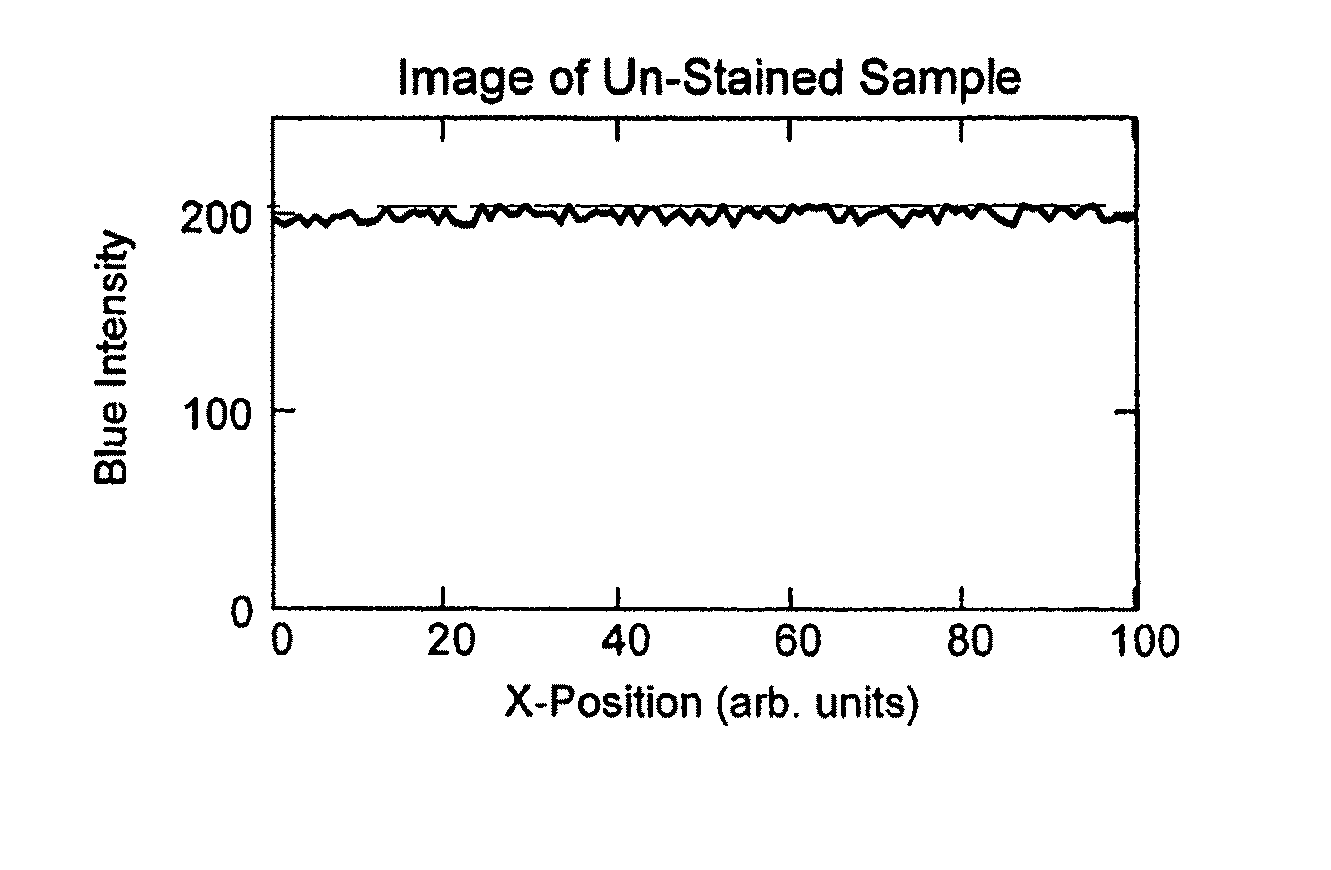
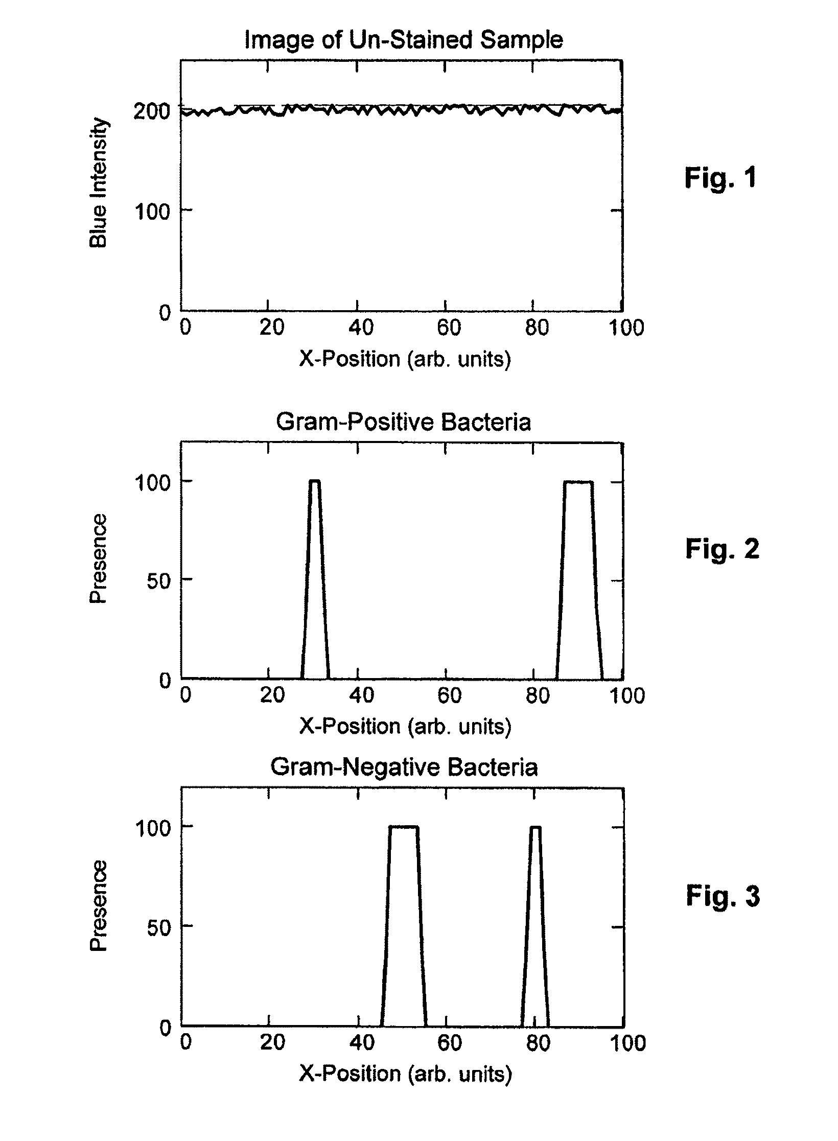
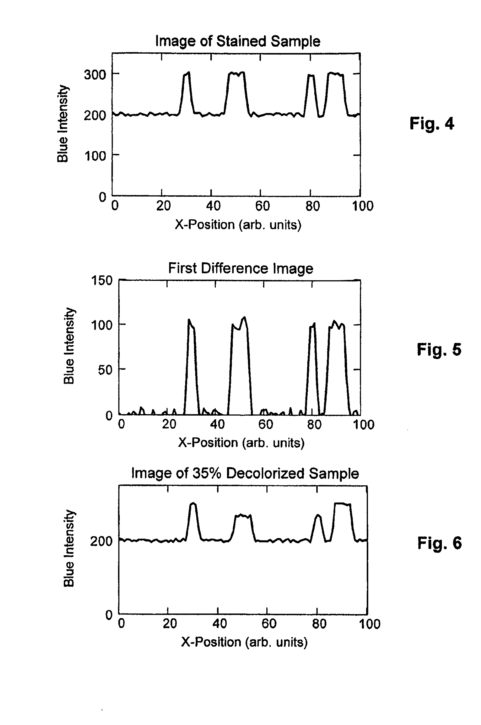
[0062]While the various method and system embodiments disclosed herein are described in the context of a Gram staining procedure, it should be understood that the various method and system embodiments described herein may also be applied to many other staining procedures that may cause optical and / or colorimetric changes in a sample. Furthermore, it should be emphasized that the exemplary images shown in FIGS. 1-29 represent only one line of a two-dimensional image. In reducing the various method and system emb...


PUM


Abstract

Various embodiments of the present invention provide methods and systems for optimally staining biological samples for analysis. In one embodiment, images of a sample are recorded before and after the application of a staining reagent and a decolorization reagent. A difference image is then generated based at least in part on a comparison of the images of the sample recorded before and after the application of the staining and decolorization reagents. Application parameters of the staining and decolorization reagents are then corrected based at least in part on the difference image such that the reagents may be optimally reapplied to generate a final image of the sample that may enable a user to better differentiate at least one target of interest in the sample. In one example, embodiments of the present invention allow for the generation of images that show only Gram-positive bacteria or only Gram-negative bacteria.

Description

CROSS REFERENCE TO RELATED APPLICATIONS[0001]This application is a national stage application filed under 35 U.S.C. 371 of International Application No. PCT / US2008 / 055471, filed Feb. 29, 2008, which claims priority from U.S. Provisional Application No. 60 / 892,736, filed Mar. 2, 2007, each of which is incorporated by reference in its entirety herein.FIELD OF THE INVENTION[0002]The present invention relates to methods and apparatus for the staining of biological material for the purpose of detecting and identifying disease-causing microorganisms. More particularly, the present invention relates to methods and apparatus for performing automated Gram stains on microscope slides.BACKGROUND OF THE INVENTION[0003]Gram staining is one of the most frequently performed procedures in modern microbiology laboratories. Such procedures are used to broadly classify bacteria as “Gram-positive” or “Gram-negative.” After affixing a bacteria-containing sample to a microscope slide, the sample is treat...

Claims


Application Information

Patent Timeline
Patent Type & Authority Patents(United States)
IPC IPC(8): G06K9/00
CPCG01N1/30G01N21/251G01N1/312
Inventor BERNDT, KLAUS W.
Owner BECTON DICKINSON & CO
Why Eureka
Try Eureka