ECG triggered heart and arterial magnetic resonance imaging

a heart and arterial magnetic resonance imaging technology, applied in the field of ecg triggered heart and arterial magnetic resonance imaging, can solve the problems of difficult to obtain precise magnetic resonance images of the heart and its connected vessels for study and comparison, and the difficulty of pooling and emptying of blood for stable imaging

Inactive Publication Date: 2014-01-07
PERINATRONICS MEDICAL SYST
View PDF23 Cites 24 Cited by
  • Summary
  • Abstract
  • Description
  • Claims
  • Application Information

AI Technical Summary

Problems solved by technology

Hearts and blood vessels are in constant motion and collecting, pooling and emptying of blood presents difficulty in stable imaging.
Consequently it is difficult to obtain precise magnetic resonance images of the heart and its connected vessels for study and comparison.

Method used


Image

  • ECG triggered heart and arterial magnetic resonance imaging
  • ECG triggered heart and arterial magnetic resonance imaging
  • ECG triggered heart and arterial magnetic resonance imaging

Examples


Embodiment Construction

Overview of Preferred Embodiment

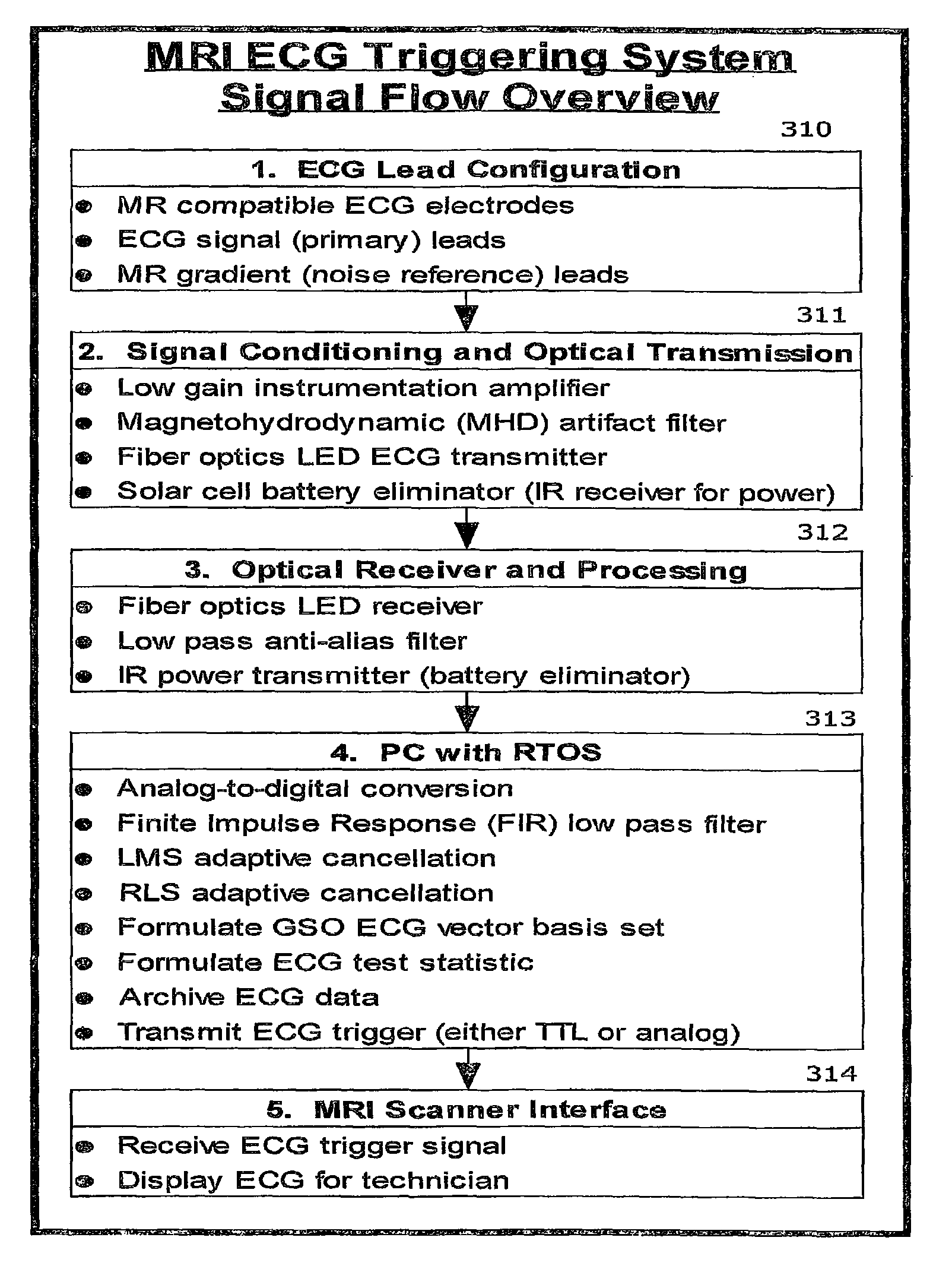
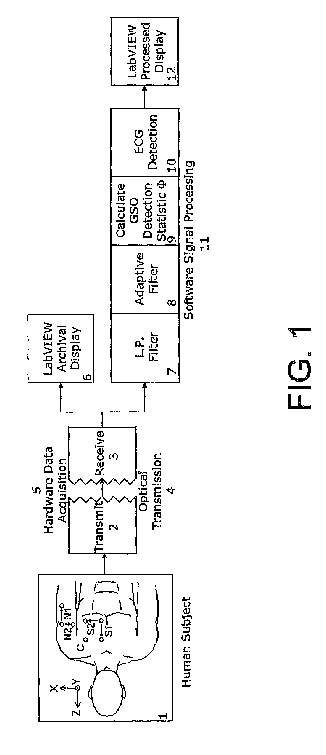
[0042]The preferred embodiment, shown in FIG. 1, consists of electrodes placed on the chest and arm of a human subject 1. A hardware data acquisition system 5 acquires the electrocardiogram and electrocardiographic noise from the chest and arm electrodes and optically transmits 2, 4 those signals out of the MRI scanner. Outside of the MRI scanner, these signals are received 3, converted back to electrical signals, and the data archived to a LabVIEW archival display computer program 6. In addition, these signals are concurrently processed by a number of software signal processing modules 11. The software signal processing consists of a low pass filter 7, one of various adaptive filters 8, a module that calculates a detection statistic using a GSO vector 9 and a module that performs ECG detection 10 and transmits a signal to the MRI to emit MR gradient pulse sequences and an RF signal to produce images. Finally, the detected electrocardiogram signal is ...


PUM


Abstract

High resistance non-metallic ECG leads are used to capture biologically generated electrical signals, and include at least one magnetic resonance noise lead to capture a noise reference signal indicative of electromagnetic noise ambient to the leads generated by magnetic resonance imaging (MRI). The noise reference signal is canceled from the captured electrical signal using an adaptive canceling noise filter to obtain a processed electrical signal indicative of the biologically generated electrical signal that causes movement in a patient's moving body part, such as the heart. A characteristic of the processing electrical signal indicative of the biologically generated electrical signal that causes the movement is detected to obtain a trigger signal, which is then transmitted to cause the MRI system to capture at least one imagine including the moving body part.

Description

[0001]This application is a continuation of U.S. patent application Ser. No. 13 / 234,762, filed Sep. 16, 2011 now abandoned, which is a continuation of U.S. patent application Ser. No. 11 / 809,876, filed on Jun. 1, 2007 now abandoned, which claims the priority benefit of U.S. Provisional Application No. 60 / 809,833, filed Jun. 1, 2006, which are hereby incorporated by reference in their entirety.GOVERNMENT LICENSE RIGHTS[0002]This invention was made with government support under Grant HL66791, awarded by the National Institutes of Health. The government has certain rights in the invention.BACKGROUND OF THE INVENTION[0003]Magnetic resonance imaging of hearts and connected blood vessels is the gold standard of determining pericardial structure, disease and function of the heart and its major connected vessels.[0004]Hearts and blood vessels are in constant motion and collecting, pooling and emptying of blood presents difficulty in stable imaging. The beating of the heart and movement of t...

Claims


Application Information

Patent Timeline
Patent Type & Authority Patents(United States)
IPC IPC(8): A61B5/055
CPCA61B5/055A61B5/0402A61B5/044A61B5/7292A61B5/318A61B5/339
Inventor FRANK, THOMAS H.BLAUMANIS, OTIS R.LA ROSA, JOSEPH A.WORD, YVETTE M.
Owner PERINATRONICS MEDICAL SYST
Why Eureka
Try Eureka