Inhibition of the interaction between 5T4 oncofoetal glycoproteins and CXC chemokine receptors as a method of identifying chemotaxis inhibitors

a technology of cxc chemokine receptor and oncofoetal glycoprotein, which is applied in the field of methods, can solve the problems of poor prognosis, significant challenge, and cancer metastasis treatment, and achieve the effect of being a useful target for treating pathological inflammation

Inactive Publication Date: 2014-05-06
CANCER RES TECH LTD
View PDF1 Cites 1 Cited by
  • Summary
  • Abstract
  • Description
  • Claims
  • Application Information

AI Technical Summary

Benefits of technology

[0012]The present inventors have identified a complex of approximately 130 kDa (as determined by PAGE) comprising the 5T4 polypeptide and a CXC chemokine receptor polypeptide (in particular the CXCR4 protein). The complex has been found to be stable in the cell membrane and was able to remain intact following non-ionic detergent solubilisation and unreduced PAGE. Accordingly, in some cases the test agent may inhibit the interaction between the 5T4 polypeptide and the CXC chemokine receptor polypeptide by inhibiting formation of the complex, destabilising the complex or enhancing the break-up of the complex.
[0020]Chemotaxis, particularly chemotaxis in response to CXCL12 or CXCL16, is an important feature of metastasis. Therefore, an inhibitor of the interaction between 5T4 and a CXC chemokine receptor may exhibit anti-cancer therapeutic activity, particularly inhibition or prevention of metastasis (e.g. spread of 5T4 +ve and CXCR4 +ve cancer cells to CXCL12-expressing sites or spread of 5T4 +ve and CXCR6 +ve cancer cells to CXCL16-expressing sites). Accordingly, the method of this aspect of the invention may further comprise an in vivo step of assessing metastasis of a cancer in a non-human animal model to which the test agent has been administered, as compared with metastasis of a cancer in a control non-human animal model to which the test agent has not been administered. The non-human animal model may be a mammalian laboratory animal species such as a rodent (e.g. mouse, rat), rabbit, cat, dog or non-human primate. In some cases the mouse may be a 5T4 null mouse (e.g. 5T4 KO mouse), which may be useful, for example, as a negative control. In some cases a 5T4-ve animal (e.g. a 5T4 null mouse) may be the recipient of a 5T4 positive and CXC chemokine receptor positive tumour (e.g. a 5T4 +ve and CXCR4 +ve tumour), e.g. an implanted tumour. In this way the effect of drug treatment may be assessed in the absence of any influence through normal tissue 5T4 / CXCR4 expression.
[0063]The antibody or fragment thereof, in accordance with this aspect of the invention, may be identified or identifiable in a method according to the first aspect of the invention. The skilled person is able to provide antibodies and antibody fragments comprising antibody binding domains that are capable of binding to a 5T4 polypeptide as defined in relation to the first aspect of the invention, are capable of binding to a CXC chemokine receptor polypeptide as defined in relation to the first aspect of the invention or are able to bind to a complex comprising a 5T4 polypeptide and a CXC chemokine receptor polypeptide (e.g. a 5T4-CXCR4 complex or a 5T4-CXCR6 complex). Such antibodies or fragments may be readily screened for the ability to inhibit the interaction of the 5T4 polypeptide and the CXC chemokine receptor polypeptide using a method in accordance with the first aspect of the invention. A suitable antibody may in some cases be isolated from a library of antibody molecules (e.g. a phage display library) using, e.g., a complex comprising a 5T4 polypeptide and a CXC chemokine receptor polypeptide as an antigen.
[0088]The method of this aspect of the invention is useful for providing clinically relevant prognostic information about a subject having ALL. Said poor prognosis may comprise: an elevated risk of having or developing extramedullary ALL; an elevated risk of relapse; an elevated risk of therapeutic failure; and / or a lower likelihood of survival.

Problems solved by technology

Treatment of cancer metastasis, the spread and growth of tumour cells to distant sites, presents a significant challenge despite the identification of a number of mediators that are implicated in the process.
Indeed, spread of cancer to distant sites is almost universally associated with poor prognosis and, in many cases, resistance to effective treatment.
Despite advances in the treatment of many cancers, provision of therapeutic agents and treatment strategies for preventing or limiting the spread of cancer cells is currently hampered by an incomplete picture of the mechanistic details of metastasis.

Method used


Image

  • Inhibition of the interaction between 5T4 oncofoetal glycoproteins and CXC chemokine receptors as a method of identifying chemotaxis inhibitors
  • Inhibition of the interaction between 5T4 oncofoetal glycoproteins and CXC chemokine receptors as a method of identifying chemotaxis inhibitors
  • Inhibition of the interaction between 5T4 oncofoetal glycoproteins and CXC chemokine receptors as a method of identifying chemotaxis inhibitors

Examples


example 1

Differentiating mES Cells Show 5T4-Dependent CXCL12 Chemotaxis

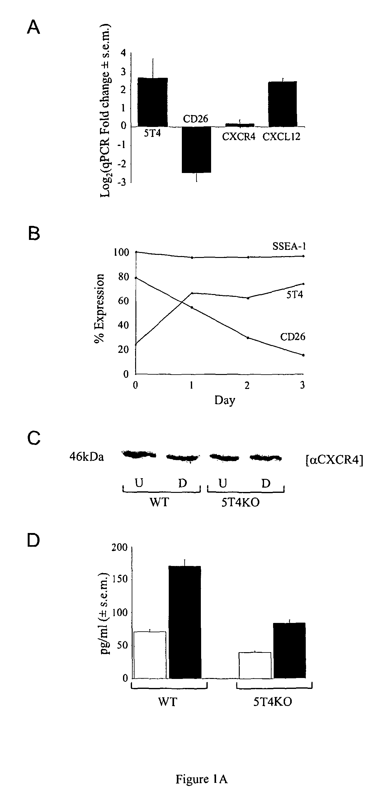
[0208]A comparative microarray of undifferentiated (5T4 −ve) and early differentiating (5T4 +ve) mES cells identified a significant downregulation of CD26, upregulation of CXCL12 but no change in the CXCR4 transcripts [30]. These data were confirmed by qPCR (FIG. 1A) and FACS analysis showed that as the ES cells differentiate cell surface expression of CD26 decreases while 5T4 increases; by contrast the pluripotent ES marker SSEA-1 did not significantly change over this time (FIG. 1B). The adherent cells were harvested for FACS analysis using trypsin-EDTA and CXCR4 is trypsin sensitive so its cell surface expression could not be accessed by this method. Western blot analysis shows there is no change in the total CXCR4 expression upon differentiation of either WT or 5T4KO ES cells (FIG. 1C). Increased CXCL12 was detected in the culture medium by ELISA after 3 days of differentiation (74±4 vs 180±9 pg / ml) (FIG. 1D). To exam...

example 2

Cellular Location of CXCR4 in Differentiating ES Cells

[0209]Following LIF withdrawal, both WT and 5T4KO ES cells undergo an EMT with cells eventually becoming dispersed with an arborized morphology. The expression and cellular localization of 5T4 and CXCR4 molecules before and after differentiation was determined by immunofluorescence of fixed cells grown on glass plates (FIG. 3A). Undifferentiated WT-ES cells are 5T4-negative with CXCR4 expression low and intracellular; following differentiation both molecules can be detected at the cell surface with some areas of co-localization. By contrast, differentiated 5T4KO ES cells show only intracellular CXCR4 expression. It is apparent that at least some 5T4 and CXCR4 molecules co-localize to lipid rafts in differentiating WT but not 5T4KO differentiating ES cells where CXCR4 remains intracellular. However, when differentiating 5T4KO ES cells are infected with RAd-m5T4, CXCR4 can be detected at the cell surface co-localized with 5T4 molec...

example 3

Role of 5T4 Expression in the CXCL12 / CXCR4 Axis in Mouse Embryo Fibroblasts

[0210]A 5T4 dependency for CXCR4-mediated chemotaxis is also apparent in MEFs as shown by: (1) a 5T4 gene dose influence on CXCL12 chemotaxis in WT, heterozygote and KO MEFS (FIG. 3a); (2) the restoration of the chemotactic response of 5T4 null MEF by RAd-m5T4 (FIG. 3b); and (3) the co-localization of some CXCR4 molecules with typical punctuate 5T4 cell surface expression in WT MEFs while 5T4 null MEFs show only intracellular CXCR4 (FIG. 3c) that can be rescued at the cell surface by RAd-m5T4 (FIG. 3d).

[0211]We next examined the role of 5T4 in the CXCL12 / CXCR4 axis by analyzing CXCL12-induced activation of key intracellular signalling effectors ERK and AKT in both WT and 5T4 null MEFs (FIG. 3e). These data demonstrate that in WT MEFs classical signal transduction pathways for the CXCL12 / CXCR4 axis are active but in the absence of 5T4 both ERK and AKT pathways are disrupted and the phosphorylation status of th...


PUM

PropertyMeasurementUnit
molecular weightsaaaaaaaaaa
molecular weightaaaaaaaaaa

Abstract

Methods and agents are disclosed based on the finding that 5T4 interacts with CXCR4 in the cell membrane to form a complex, and that the 5T4 transmembrane region is involved in the promotion of CXCR4 membrane expression and chemotactic response.

Description

FIELD OF THE INVENTION[0001]The present invention relates to methods, particularly methods of screening test agents, methods of treatment and methods of patient stratification, involving a glycoprotein that is involved in cancer metastasis, and to products for use in such methods.BACKGROUND TO THE INVENTION[0002]Treatment of cancer metastasis, the spread and growth of tumour cells to distant sites, presents a significant challenge despite the identification of a number of mediators that are implicated in the process. Indeed, spread of cancer to distant sites is almost universally associated with poor prognosis and, in many cases, resistance to effective treatment. For example, in childhood acute lymphoblastic leukaemia (ALL) certain high-risk cytogenetic subtypes not only have higher relapse rate but are also more likely to relapse in extramedullary sites such as the CNS. Methods for identification of tumour subtypes that are prone to spread and / or relapse are currently sub-optimal....

Claims


Application Information

Patent Timeline
Patent Type & Authority Patents(United States)
IPC IPC(8): G01N33/567G01N33/574
CPCG01N2800/52G01N33/574C12N2799/022G01N2800/54G01N2500/02G01N33/57426C07K2317/92G01N2333/471C12N2799/027C07K2316/96C07K16/32G01N33/6863C07K16/2866C07K16/30C07K2317/31G01N33/57492C07K2317/76A61P1/00A61P1/16A61P19/02A61P29/00A61P35/04A61P9/10
Inventor STERN, PETER L.SAHA, VASKAR
Owner CANCER RES TECH LTD
Why Eureka
Try Eureka