Method for minimally invasive tendon sheath release using device with hemi-cannula

a technology of hemi-cannula and tendon, which is applied in the field of devices and methods for releasing the tendon sheath, can solve the problems of causing pain, causing the tendon to swell, and causing the nodule to form, and achieves the effects of less surrounding tissue, less surrounding tissue dissection, and less pulling

Active Publication Date: 2014-07-08
SONICSURG INNOVATIONS
View PDF33 Cites 30 Cited by
  • Summary
  • Abstract
  • Description
  • Claims
  • Application Information

AI Technical Summary

Benefits of technology

[0007]Generally speaking, the claimed invention is a precisely guided scalpel device having a protective cover over the blade that allows a surgeon to perform a tendon sheath release procedure safely and quickly in an office. The procedure is performed through an incision about 90 to 95% smaller than a conventional incision while at the same time allowing for minimal dissection of surrounding tissue and a more precise release of the pulley. Probes at the tip of the device allow the user to ensure that the device is appropriately positioned under the pulley and, when this is confirmed under fluoroscopy or ultrasound, the protective cover is engaged and a cutting blade can be deployed to safely sever the pulley, thereby disturbing much less surrounding tissue than a conventional operation.

Problems solved by technology

The constant irritation from the tendon repeatedly sliding through the pulley causes the tendon to swell in this area and create the nodule.
Pain usually occurs when the finger is bent and straightened.
If the nodule becomes too large it may pass under the ligament and become stuck at the near edge.

Method used


Image

  • Method for minimally invasive tendon sheath release using device with hemi-cannula
  • Method for minimally invasive tendon sheath release using device with hemi-cannula
  • Method for minimally invasive tendon sheath release using device with hemi-cannula

Examples


Embodiment Construction

[0038]In the following detailed description of the preferred embodiments, reference is made to the accompanying drawings, which form a part hereof, and within which are shown by way of illustration specific embodiments by which the invention may be practiced. It is to be understood that other embodiments by which the invention may be practiced. It is to be understood that other embodiments may be utilized and structural changes may be made without departing from the scope of the invention.

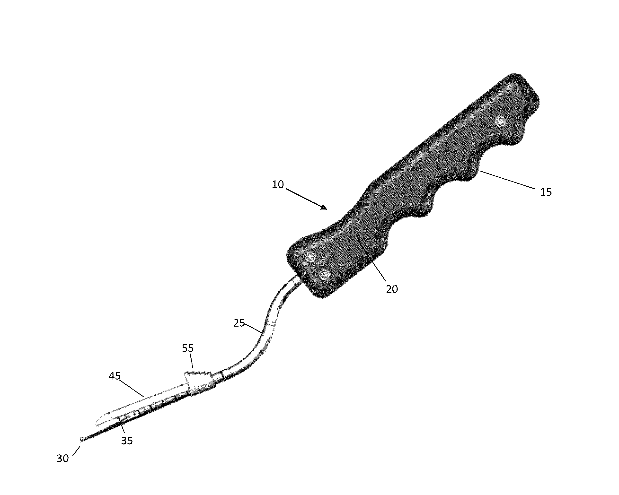
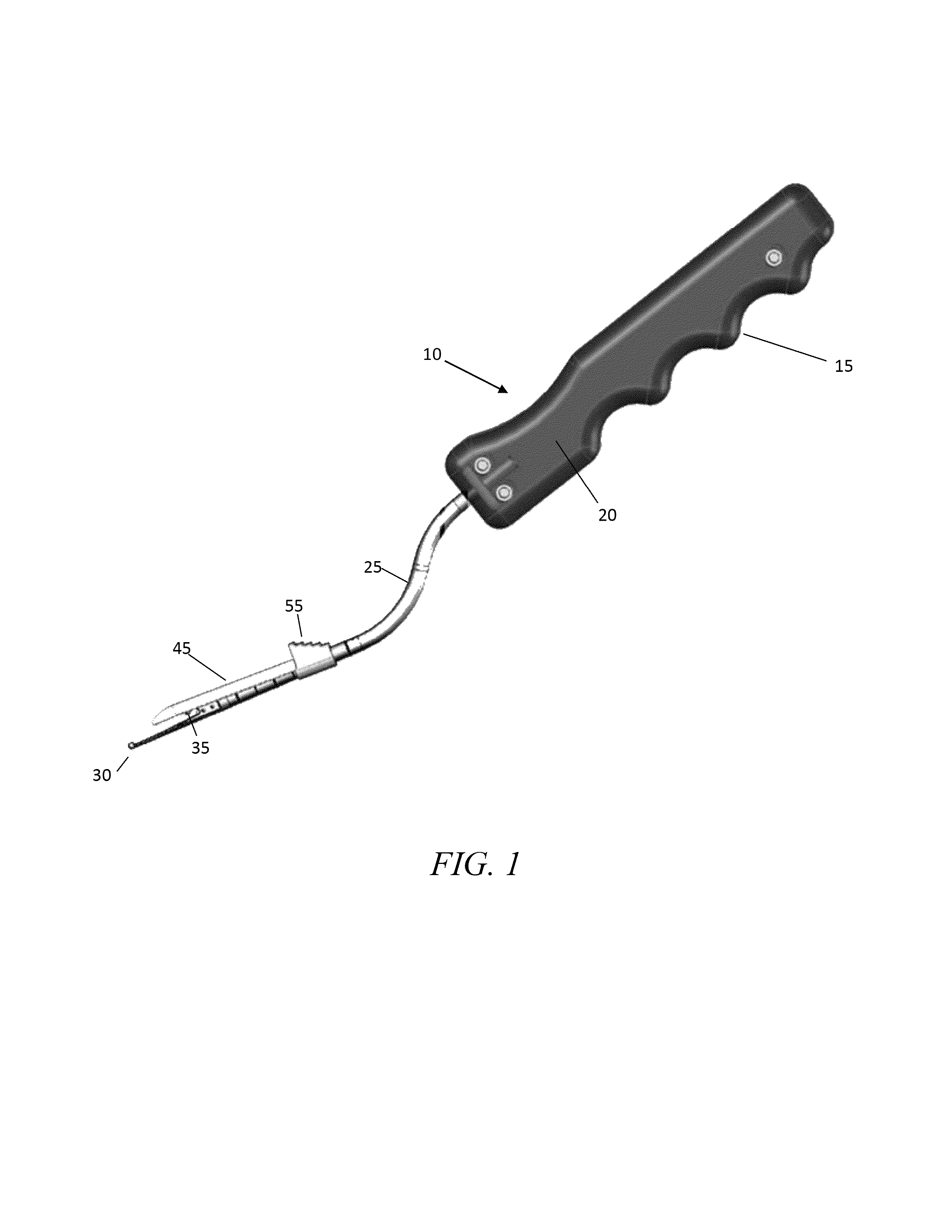
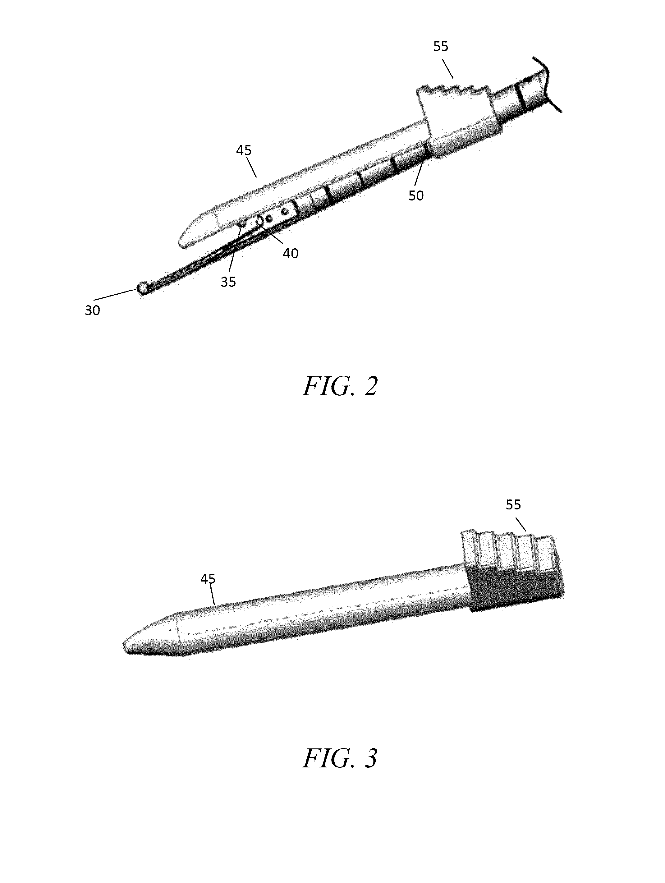
[0039]Generally, device 10 is comprised of handle 15, cavity 20, sheath 25, guide probe 30, dorsal outrigger guide probe 35, cutting blade 40 and hemi-cannula 45. Handle 15, as shown in FIGS. 1, 5, 8, 9, 12-15, and 17, includes a knurled rigid structure having dimensions of about 2.0 cm diameter and about 7.0 cm in length. Handle may be constructed of a rigid material including, but not limited to, a plastic, thermoplastic, acrylic, or metal. Handle 15 may be constructed of any shape but is prefera...


PUM


Abstract

A method for minimally invasive tendon sheath release is presented herein. The method enables a surgeon to cut (“open”) a pulley that is obstructing a nodule and keeping a tendon from sliding smoothly. A guide probe of the device is inserted through a small incision and is used to find the edge of the pulley. Once found, the probe is guided to an end of the pulley. Proper placement of the device may be confirmed through the use of ultrasound. After proper position is assured, a hemi-cannula is moved over the guide probe to isolate the pulley from the surrounding tissue. A cutting blade is then used to sever the pulley. A dilation device may be inserted prior to the insertion of the spherical tipped guide probe to dilate the area.

Description

CROSS REFERENCE TO RELATED APPLICATION[0001]This application is a divisional of and claims priority to currently pending U.S. patent application Ser. No. 13 / 273,502, entitled “Device for Minimally Invasive Tendon Sheath Release”, filed on Oct. 14, 2011, which is a continuation in part and claims priority to U.S. patent application Ser. No. 12 / 896,088, entitled “Method for Minimally Invasive Tendon Sheath Release”, filed on Oct. 1, 2010 which claims priority to U.S. Provisional Patent Application No. 61 / 251,957, entitled “Device for Minimally Invasive Tendon Sheath Release,” filed on Oct. 15, 2009, the contents of each of which are hereby incorporated by reference.FIELD OF THE INVENTION[0002]This invention relates to a novel device and method for minimally invasive tendon sheath release. More particularly, it relates to a device and method that allows a surgeon to sever a pulley that is obstructing a nodule without damaging any surrounding tissue.BACKGROUND OF THE INVENTION[0003]Tend...

Claims


Application Information

Patent Timeline
Patent Type & Authority Patents(United States)
IPC IPC(8): A61B17/32
CPCA61B17/320036A61B34/20A61B2090/08021A61B17/320016
Inventor JURBALA, BRIAN, MICHAEL
Owner SONICSURG INNOVATIONS
Why Eureka
Try Eureka