Methods and apparatuses for analyzing biological samples by mass spectrometry

a mass spectrometry and biological sample technology, applied in the field of medical imaging, analysis, monitoring and diagnostics, can solve the problems of non-specific and inaccurate techniques, conventional technology does not employ maldi ms techniques to effectively analyze proteins in clinical samples, and conventional technology does not use maldi ms techniques to perform the effect of analyzing proteins

Inactive Publication Date: 2014-07-15
VANDERBILT UNIV
View PDF18 Cites 32 Cited by
  • Summary
  • Abstract
  • Description
  • Claims
  • Application Information

AI Technical Summary

Problems solved by technology

Such techniques, however, are, at times, non-specific and inaccurate.
More importantly, conventional technology does not involve techniques that effectively employ the capability of MALDI MS to analyze and effectively depict a quantity of molecules of interest with a specific molecular mass or within a selected molecular mass window or as a function of their position on the test sample.
Further, conventional technology does not employ MALDI MS techniques to effectively analyze proteins in clinical samples.
Still further, conventional technology does not use MALDI MS techniques to perform direct profiling of diseased tissue, which in turn may be used for the assessment of disease classification, development and treatment.

Method used


Image

  • Methods and apparatuses for analyzing biological samples by mass spectrometry
  • Methods and apparatuses for analyzing biological samples by mass spectrometry
  • Methods and apparatuses for analyzing biological samples by mass spectrometry

Examples


example 1

General Methodology

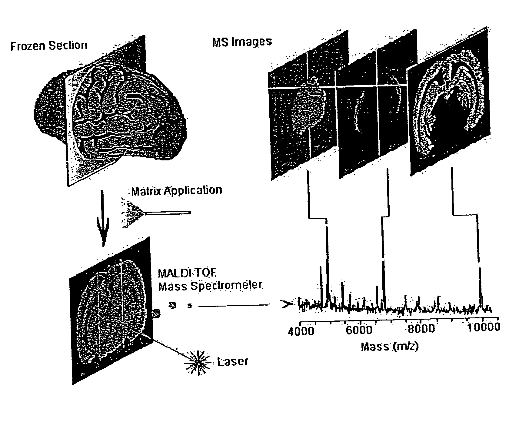
[0061]FIG. 1 illustrates methodology suitable for embodiments of this disclosure. The illustrated methodology of FIG. 1 was designed for the spatial analysis of tissue by MALDI mass spectrometry. In this embodiment, frozen sections are mounted on a metal plate, coated with an UV-absorbing matrix and placed in a mass spectrometer. A pulsed UV laser desorbs and ionizes analytes from the tissue and their m / z values are determined using an instrument such as a time-of-flight analyzer. From a raster over the tissue and measurement of the peak intensities over several spots (which may be thousands of spots), mass spectrometric images may be generated at specific molecular weight values.

[0062]For the molecular image analysis, tissue samples can be prepared using several protocols: (a) direct analysis of fresh frozen sections, individual cells or clusters of cells isolated by laser-capture microdissection or other cell isolation procedure, or (b) contact blotting of a tis...

example 2

Application to Mammalian Tissue

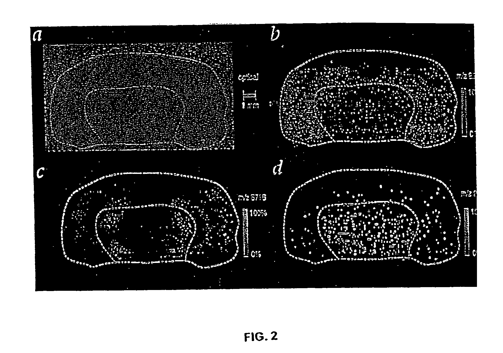
[0063]The inventor and assistants have used imaging MS to study normal tissue sections from mouse brain and human brain tumor xenograph sections. These samples contained well-defined regions, many of which had subsets of proteins and peptides in a unique distribution or array. The bilateral symmetry of the brain provided an internal confirmation of the localized distribution of proteins and the homogeneity of the prepared tissue sections. An optical image of the normal mouse brain section fixed on a metal plate and coated with matrix is shown in FIG. 2A. The inventor and assistants scanned the section by acquiring 170×90 spots with a spot-to-spot center distance of 100 μm in each direction. Ions occurring in 82 different mass ranges were recorded, and images were created by integrating the peak areas and plotting the relative values using a color scale. For specific molecular images, data was acquired in a window delimited by two mass-to-charge (m / z) u...

example 3

Preparation of Tumor-Bearing Tissue for Molecular Imaging

[0064]Tumor-bearing tissues were generated by subcutaneous implantation of human glioblastoma cells (D54) into the hind limb of a nude mouse. After tumors grew to about 1 cm in diameter, they were surgically removed them from the mouse and immediately frozen using liquid nitrogen. For image analysis, the inventor cut the tumor tissue using a microtome in 12-μm thick sections orthogonal to the point of attachment to normal tissue. Frozen sections were processed following the protocol described above before image analysis by MS.

[0065]The optical image of a frozen human glioblastoma section taken immediately following mass spectrometric imaging is shown in FIG. 3A. The orientation in FIG. 3A is such that the actively growing area of the tumor is at the top of the figure, and the point where the tumor was attached to the healthy tissue at the bottom. The fine line (cross-hatched) pattern on the optical image was produced by laser ...


PUM

PropertyMeasurementUnit
diameteraaaaaaaaaa
distanceaaaaaaaaaa
diameteraaaaaaaaaa

Abstract

Methods and apparatuses for analyzing proteins and other biological materials and xenobiotics within a sample. A specimen is generated, which may include an energy absorbent matrix. The specimen is struck with laser beams such that the specimen releases proteins. The atomic mass of the released proteins over a range of atomic masses is measured. An atomic mass window of interest within the range of atomic masses is analyzed to determine the spatial arrangement of specific proteins within the sample, and those specific proteins are identified as a function of the spatial arrangement. By analyzing the proteins, one may monitor and classify disease within a sample.

Description

[0001]This is a continuation of U.S. patent application Ser. No. 10 / 178,067, filed Jun. 21, 2002, now U.S. Pat. No. 6,756,586, which claims priority to U.S. Provisional Patent Application Ser. No. 60 / 329,719, filed Oct. 15, 2001. The entire contents of each of the foregoing applications is incorporated by reference.[0002]Aspects of this invention were made with government support of the National Institute of Health, grant number GM58008. Accordingly, the government may have certain rights in this invention.BACKGROUND OF THE INVENTION[0003]1. Field of the Invention[0004]The present invention relates generally to the fields of medical imaging, analysis, monitoring and diagnostics. More particularly, it provides apparatuses and methods for analyzing proteins in samples. Even more particularly, it may be used in the direct profiling of diseased tissue by mass spectrometry; this, in turn, may be used for the assessment of disease classification, development and treatment.[0005]2. Descrip...

Claims


Application Information

Patent Timeline
Patent Type & Authority Patents(United States)
IPC IPC(8): G01N24/00G01N27/62G01N27/64G01N33/68
CPCG01N33/6848G01N2800/52Y10T436/24
Inventor CAPRIOLI, RICHARD
Owner VANDERBILT UNIV
Why Eureka
Try Eureka