Magnetic resonance imaging device

a magnetic resonance imaging and magnetic field technology, applied in the field of magnetic resonance imaging devices, can solve the problems of degrading image resolution, reducing the number of sampling points, prolonging the measurement time, etc., and achieves favorable water/fat separation and ease of measurement restrictions.

Active Publication Date: 2014-07-22
FUJIFILM HEALTHCARE CORP
View PDF6 Cites 3 Cited by
  • Summary
  • Abstract
  • Description
  • Claims
  • Application Information

AI Technical Summary

Benefits of technology

[0034]According to the present invention, in a method of obtaining images with different echo times for separation of water and fats images, the restrictions concerning the measurement are eased, and favorable water / fat separation can be realized. Furthermore, it becomes possible to determine which is each of the obtained two images, a water image or a fat image.

Problems solved by technology

In the method of Non-patent document 2 mentioned above, it is necessary to carry out the measurement three times, and therefore it has a problem that the measurement time is prolonged.
Because dt is narrow at a magnetic field strength of 3.0 teslas or 1.5 teslas, the method suffers from restrictions concerning measurement, for example, the measurement band must be widened, and number of sampling points must be decreased.
If the number of sampling points is decreased, the spatial resolution of the image is degraded, and thus it becomes difficult to diagnose minute pathological lesions.

Method used


Image

  • Magnetic resonance imaging device
  • Magnetic resonance imaging device
  • Magnetic resonance imaging device

Examples


Embodiment Construction

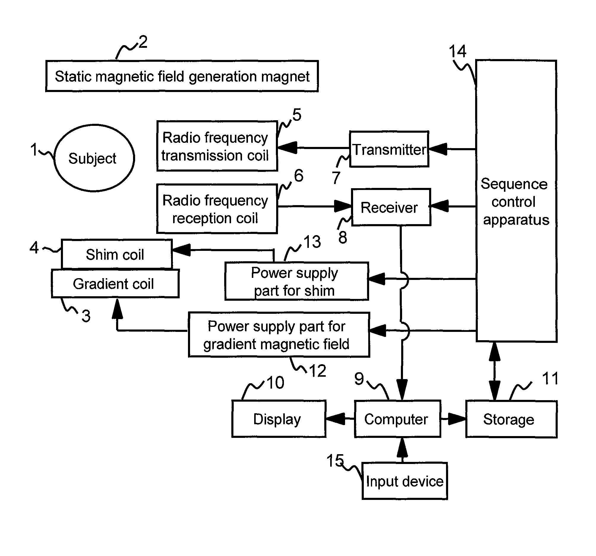
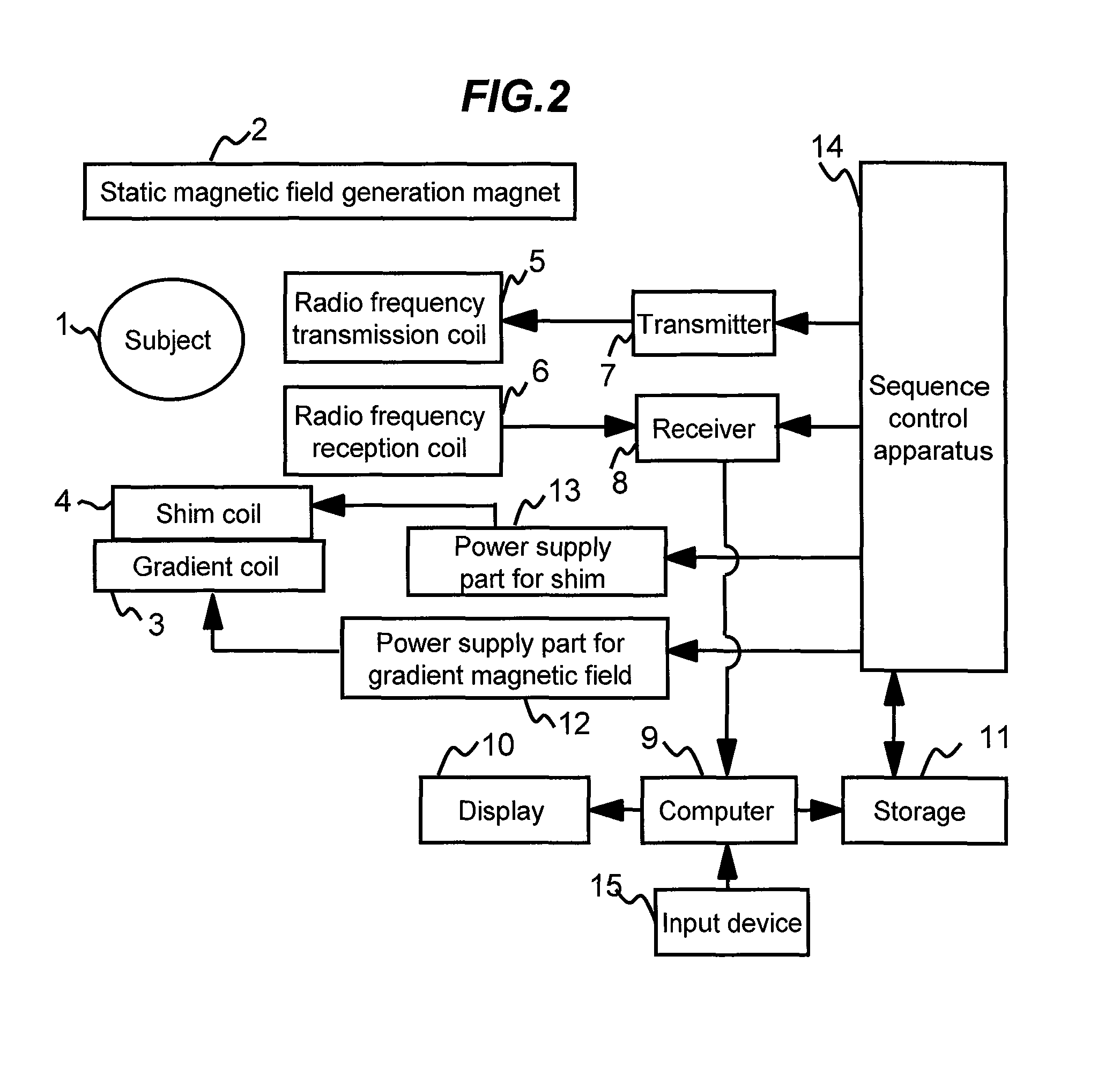
[0054]Hereafter, an embodiment of the MRI device of the present invention will be explained with reference to the drawings.

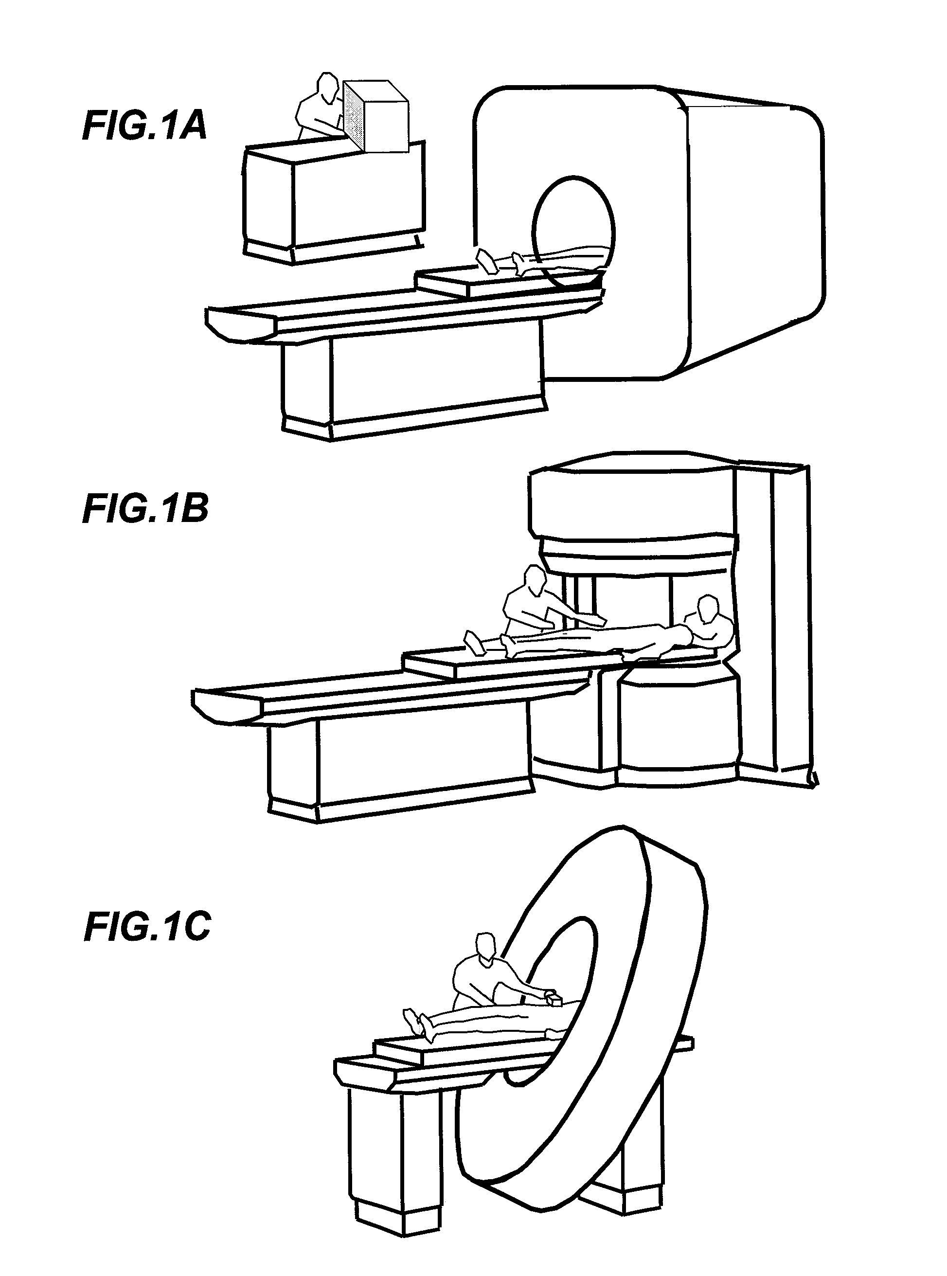
[0055]FIG. 1A to FIG. 1C show total configurations and external views of magnetic resonance imaging devices corresponding to embodiments of the present invention. FIG. 1A shows an MRI device of the horizontal magnetic field type utilizing a tunnel-shaped magnet that generates a static magnetic field with a solenoid coil. FIG. 1B shows an MRI device of the vertical magnetic field type utilizing a hamburger type (open type) magnet having separated upper and lower magnets in order to increase spaciousness. Further, FIG. 1C shows a tunnel type MRI device similar to that of FIG. 1A, but depth of the magnet is shortened and the magnet is leaned to increase spaciousness. The present invention can be applied to MRI devices of known structures including MRI devices of these configurations, but the present invention is not limited to these, and can be applied regardless o...


PUM


Abstract

Water / fat separation imaging is performed by an MRI device, even if the measurement is not performed with such echo times that the phases of water and fat signals become in-phase or out-of-phase. A determination is made as to which of two obtained separation images is a water image or a fat image. Two water / fat ratio maps are calculated from two original images obtained with such two echo times that phase differences of water and fat signals do not become positive and negative values, and do not becomes integral multiples of π. Two phase maps are calculated from the two water / fat ratio maps and are combined to calculate two minimum phase difference maps showing minimum spatial phase difference. From dispersion in differential maps obtained by spatially differentiating the minimum phase difference maps, a correct minimum phase difference map is determined, and is used to perform phase correction of the original image.

Description

TECHNICAL FIELD[0001]The present invention relates to a magnetic resonance imaging device (henceforth referred to as MRI device), and in particular, it realizes favorable water / fat separation in water / fat separation imaging, even if the measurement is not performed with such an echo time that the phases of water and fat signals become in-phase or out-of-phase. The present invention further relates to a device and a method enabling distinction of obtained two separate images, i.e., to determine which is each of them, a water image or a fat image.BACKGROUND ART[0002]The currently most widely used nuclide for imaging in clinical MRI is hydrogen nucleus (proton), which is the major constituent of subjects.[0003]MRI realizes two-dimensional or three-dimensional imaging of forms or functions of human head, abdominal part, limbs, and so forth by imaging spatial distribution of proton density or relaxation information of magnetic resonance signals.[0004]When a human body is an object of the...

Claims


Application Information

Patent Timeline
Patent Type & Authority Patents(United States)
IPC IPC(8): G06K9/00
CPCG01R33/4828A61B5/055A61B5/4869A61B5/4872
Inventor SHIRAI, TORUBITO, YOSHITAKA
Owner FUJIFILM HEALTHCARE CORP
Why Eureka
Try Eureka