
Automated pneumothorax detection

a pneumothorax and automatic technology, applied in the field of automatic detection of lung related ailments or conditions, can solve problems such as hemodynamic instability and/or death

Active Publication Date: 2014-12-16
THE JOHN HOPKINS UNIV SCHOOL OF MEDICINE
View PDF19 Cites 12 Cited by
  • Summary
  • Abstract
  • Description
  • Claims
  • Application Information

AI Technical Summary

Problems solved by technology

The severity of the collapse of one, or sometimes both, lungs determines the oxygenation and ventilation impairment for the patient, which can lead to hemodynamic instability and / or death.
The symptoms of pneumothorax are often vague and inconclusive, so that medical imaging is required to achieve an accurate diagnosis of the condition.

Method used


Image

  • Automated pneumothorax detection
  • Automated pneumothorax detection
  • Automated pneumothorax detection

Examples


Embodiment Construction

[0017]Some example embodiments now will be described more fully hereinafter with reference to the accompanying drawings, in which some, but not all example embodiments are shown. Indeed, the examples described and pictured herein should not be construed as being limiting as to the scope, applicability or configuration of the present disclosure. Rather, these example embodiments are provided so that this disclosure will satisfy applicable legal requirements. Like reference numerals refer to like elements throughout.

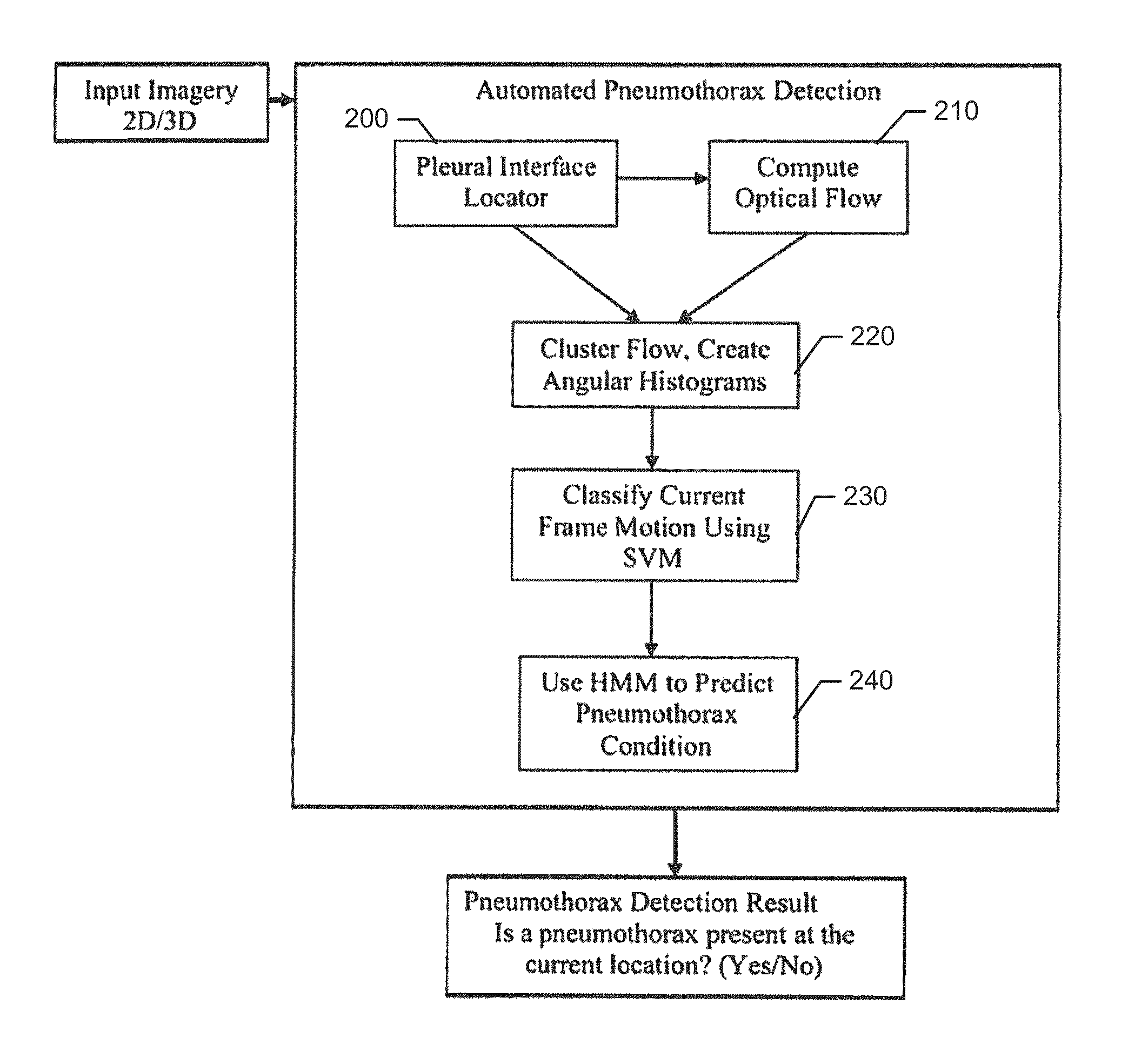
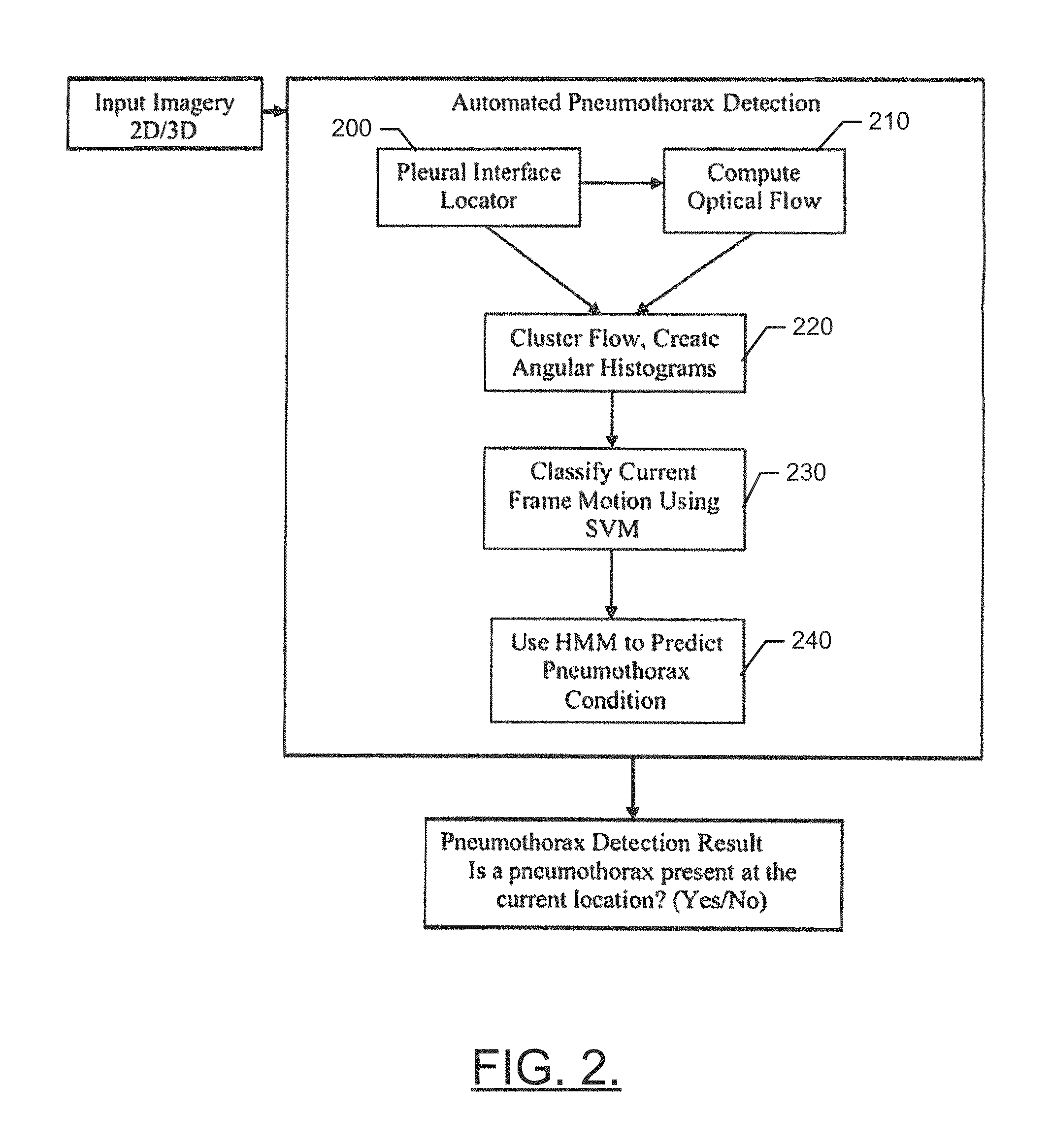
[0018]As indicated above, some example embodiments may enable the provision of a mechanism by which to diagnose pneumothorax automatically on the basis of machine executed analysis of image data of lungs. In some cases, the image data may be one-dimensional, two-dimensional or three-dimensional video imagery that may be obtained by time varying imaging modalities such as ultrasound (including Doppler ultrasound), CT or cine-MRI. The image data may be analyzed to identify o...


PUM


Abstract

A method of determining the presence of a pneumothorax includes obtaining a series of frames of image data relating to a region of interest including a pleural interface of a lung. The image data includes at least a first frame and a second frame. The method further includes identifying, via processing circuitry, the pleural interface in at least the first frame and the second frame, determining, based on computing optical flow between the first and second frames, a pleural sliding classification of the image data at the pleural interface, and determining whether a pneumothorax is present in the pleural interface based on the pleural sliding classification.

Description

CROSS-REFERENCE TO RELATED APPLICATION[0001]This application claims priority to and the benefit of U.S. Provisional Application Ser. No. 61 / 592,285 filed on Jan. 30, 2012, the entire contents of which are hereby incorporated herein by reference.TECHNICAL FIELD[0002]Example embodiments of the present disclosure generally relate to automatic detection of lung related ailments or conditions, and more specifically relate to employment of automated image analysis to facilitate detection of pneumothoraces.BACKGROUND[0003]A pneumothorax is an abnormal lung condition that is characterized by the collection of air in the pleural space separating the lung from the chest wall. Pneumothoraces may occur in one or both lungs and cause a partial or complete collapse of the affected lung or lungs. The severity of the collapse of one, or sometimes both, lungs determines the oxygenation and ventilation impairment for the patient, which can lead to hemodynamic instability and / or death.[0004]The sympto...

Claims


Application Information

Patent Timeline
Patent Type & Authority Patents(United States)
IPC IPC(8): G06K9/46A61B5/00G06T7/20G06K9/62G06T7/00A61B6/00G06V10/50G06V10/764
CPCG06T2207/20061G06K9/6297G06T2207/10072G06K9/6269G06T7/2033A61B5/0077G06T2207/10132G06K9/4642G06T2207/30061G06T7/0016G06T2207/10016A61B8/5223A61B5/7282A61B5/0873A61B8/08A61B6/5217A61B5/055A61B6/032A61B5/7445A61B5/7264G06T7/246G06V10/50G06V10/85G06V10/764G06F18/295G06F18/2411
Inventor BURLINA, PHILIPPE M.MUKHERJEE, RYAN N.
Owner THE JOHN HOPKINS UNIV SCHOOL OF MEDICINE
Learn More