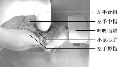
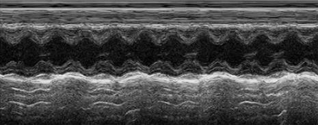
[0003] The methods for making the mouse myocardial infarction model include
drug method, minimally invasive method, electrical stimulation method,
electrocoagulation cautery method, freezing method, left ventricular anterior descending
artery ligation method; the
drug method is intravenous infusion of isoproterenol or Intraperitoneally injected into mice, the coronary
artery spasm is induced by the
drug to promote myocardial infarction, but due to the wide range of effects of the drug, the
dose used, and the individual's sensitivity to the drug, the infarction caused by this method is often more diffuse. , the
pathological change process of the prepared myocardial infarction model is also different from the
pathological change process of myocardial infarction, which is not convenient for the later evaluation of myocardial infarction size, and the reliability of this method is relatively low and has been seldom used; The innovative method is to inject the
gelatin sponge embolus into the distal end of the left anterior descending coronary artery with diatrizoate
meglumine through the
angiography tube on the basis of the
coronary angiography of the animals, so as to complete the
embolization of the model. The interventional operation is extremely difficult, and the experimental cost is high, so this method is not suitable for use; the electrical stimulation method is to
cut open the
thorax to
expose the heart with the assistance of an animal ventilator, and use a controllable micro-current to stimulate the vascular
adventitia, resulting in damage to the vascular intima. Activation of platelets induces the coagulation process to form a
thrombus leading to myocardial infarction. Because the
thrombus produced is poorly controllable and causes
thrombus to occur in other parts, there are many complications. The
electrocoagulation cautery method is to
cut open the chest to
expose the heart with the assistance of an animal ventilator. The electrocautery
electrode of the high-frequency knife cauterizes the deep part of the anterior descending
branch of the left
ventricle to cause myocardial infarction. This method is due to myocardial injury and
necrosis caused by ultra-high temperature conditions, not myocardial
ischemic necrosis. The modeling process is consistent with myocardial infarction. There are great differences in the natural
pathogenesis of infarction. The inflammatory reaction in the cauterized area is relatively strong, and the post-treatment steps are complicated. Because the thoracic opening of the mouse is small, the
heartbeat of the mouse is fast, and the operation is difficult; the freezing method is assisted by an animal ventilator. ,
cut open the
thorax to
expose the heart, put the
metal rod in
liquid nitrogen for a few minutes and take it out, the top of the
metal rod touches the anterior descending
branch of the mouse, causing the freezing injury of the anterior descending
branch, blocking the
blood flow and forming myocardial infarction, this method requires
thoracotomy The wound is large and the inflammatory reaction is strong. In order to achieve the freezing effect, it is often necessary to freeze repeatedly, which takes a long time. The degree of freezing and the range of freezing are different, resulting in poor consistency of the myocardial infarction model. out of order
[0004] The
ligation of the left ventricular anterior descending artery is the most commonly used method for modeling myocardial infarction in current experiments. This method can directly cause left ventricular anterior descending artery blockage, the test period is short, and the damage to the myocardium after infarction can be observed. The existing operation The method is to intubate the mouse through the orotrachea after anesthesia, mechanically assist ventilation, and ligate the anterior descending branch of the left
ventricle under
visual inspection after
thoracotomy. The tightness between the tube and the trachea wall is poor, the ventilation efficiency is low, and the operation takes a long time. After the
thoracotomy of the mouse, in order to ensure a
high survival rate of the follow-up mice, the chest opening is small and the operating
field of view is small. The number of times the
mouse heart beats in the
thoracic cavity exceeds 400 times Every minute, the accuracy of
needle insertion to ligate the left anterior descending branch of the left
ventricle is low, time-consuming and laborious, and it is very easy to puncture into the ventricle and atrium, causing
massive bleeding and death. During
mechanical ventilation, the expansion of the lungs on both sides can reach the
surgical operation area of the chest opening. Needling can easily cause
lung injury and lead to subsequent
pneumothorax death in mice. This method of
ligation of the anterior descending branch of the left ventricle under
direct vision after thoracotomy using a ventilator to assist
breathing completes the production of a mouse model of myocardial infarction The time required is 40-90 minutes. The use of intramuscular or
intraperitoneal injection of anesthesia requires the anesthesia of the mouse to last for at least 90 minutes. The use of anesthesia injection for a long time keeps the mouse in an anesthesia state, which seriously inhibits the mouse
central nervous system and affects the coronary artery. The state of
arterial blood flow, unable to wake up immediately after the operation, and the body temperature is too low, eventually leading to the death of mice. When the myocardial infarction model is made by this method, the success rate of myocardial infarction is low and the
mortality rate of mice is high, which seriously affects the speed and efficiency of
experimental research. ; In order to solve this problem, there is an urgent need for a method for quickly making a mouse myocardial infarction model, which can optimize the operation process, reduce
operation time, improve the success rate of the myocardial infarction model, and reduce the
mortality rate of mice. Some researchers used the heart
extrusion method to make Mouse myocardial infarction model, but due to lack of mastery of the operation method and reasonable optimization of the production process, the success rate of the mouse myocardial infarction model is low and the
mortality rate is high


