
Lung volume-reducing elastic implant and instrument

an elastic implant and volume-reducing technology, applied in the field of interventional therapy, can solve the problems of high operative cost, limited cure effect, significant mental and physical pain, etc., and achieve the effect of avoiding the occurrence of pneumothorax in the delivery process, facilitating the pre-threading of guidewires, and reducing the occurrence rate of pneumothorax

Active Publication Date: 2018-05-17
SHENZHEN LIFETECH RESPIRATION SCI CO LTD
View PDF1 Cites 4 Cited by
  • Summary
  • Abstract
  • Description
  • Claims
  • Application Information

AI Technical Summary

Benefits of technology

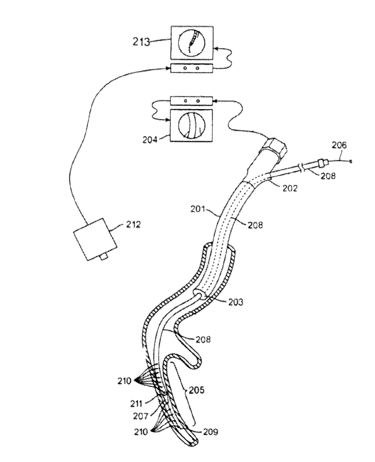
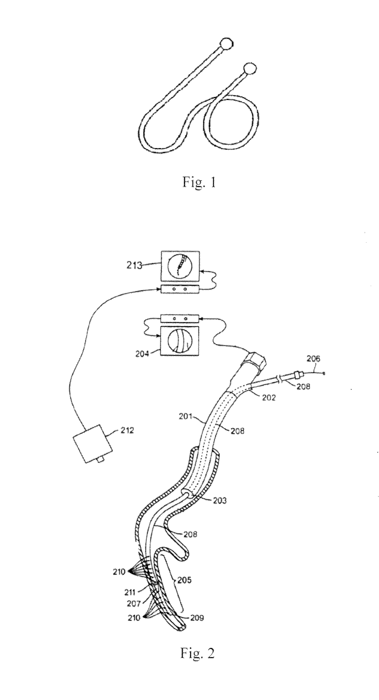
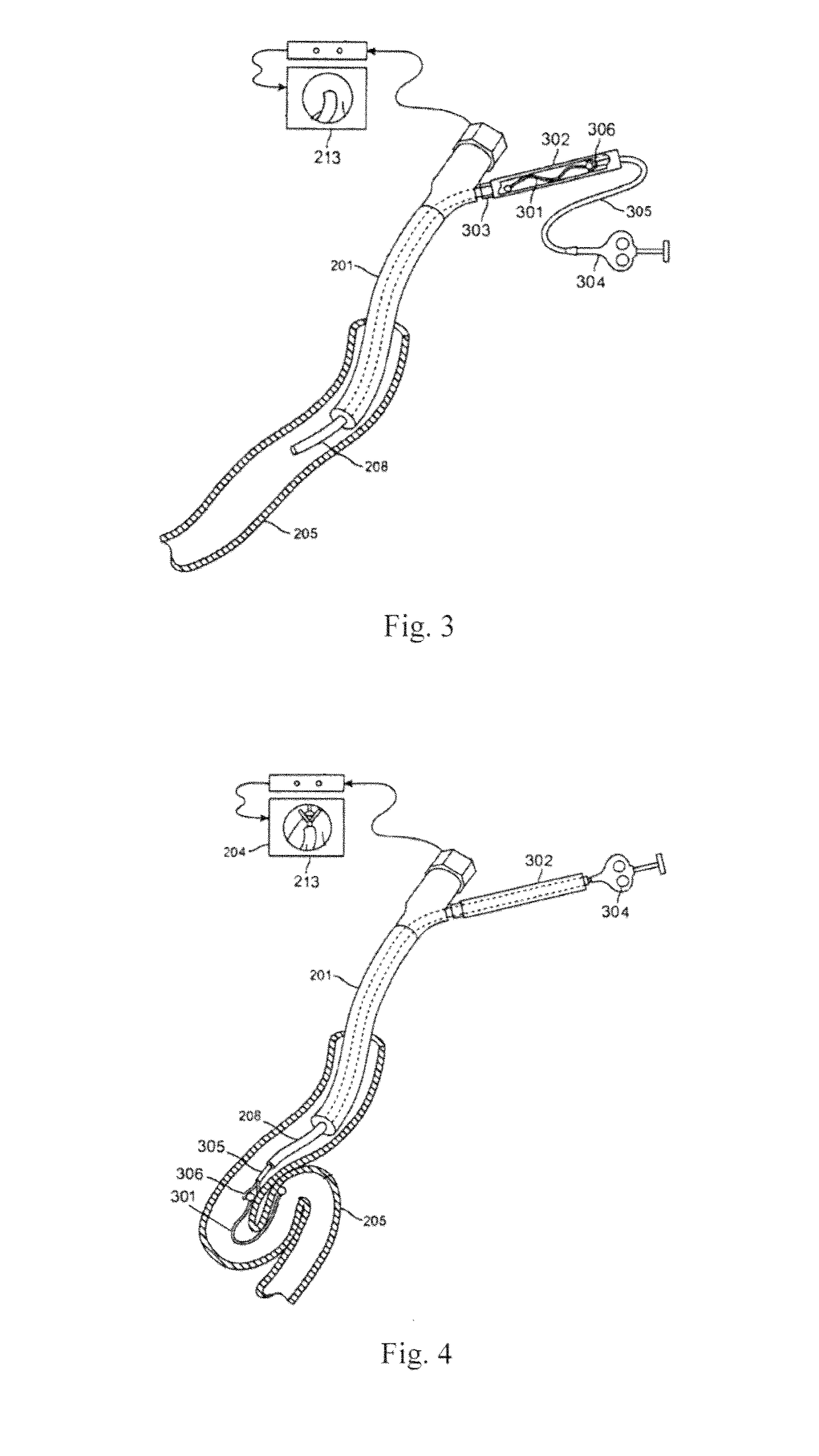
The present invention provides an implant that can be directly delivered using a guidewire instead of a delivery sheath, which reduces injury to the bronchial inner wall and reduces the occurrence of pneumothorax. Additionally, a lung volume-reducing instrument can be implanted at the tail end of a lung bypass or small-diameter tracheae according to actual requirements, combining the channel establishing process and the implant implanting operation process to make the operation more convenient, reduce the operation time, and achieve better treatment effects.

Problems solved by technology

Traditional emphysema medical treatment comprises oxygen inhalation, prevention of pulmonary infection, bronchia spasmolysis, etc. but curative effect is extremely limited.
While the surgical treatment of emphysema is given priority to lung volume-reducing surgery, and there are also relatively large amounts of limitations, such as strict indications accompanied with relatively large amounts of complications, anesthesia and complications associated therewith, the curative effect is difficult to predict before surgery, any undesirable curative effect caused by too much or too little removal cannot be compensated after surgery, and the operative cost is high, and mental and physical pain is significant.
Additionally, some patients with poor lung function often cannot tolerate the surgeries, therefore possessing high postoperative mortality, which limits the application of surgery.
Owing to the fact that the target area residual gas and sputum fails to be discharged effectively and actively, a unidirectional valve has poor clinical index, so the U.S. FDA has not approved this device.
Additionally, the effectiveness of unidirectional valve treatment is further restricted by the technical difficulty of collateral ventilation and accurate positioning on different anatomical structures.
The problem of postoperative inflammation has also not been adequately solved, due to the fact that the emphysema area is completely blocked by the xanthan gum.
In addition, the water vapor is subjected to thermal ablation, which causes postoperative inflammation because of its damage to the original tissue structure of the emphysema area.
1. The elastic coil made from the prior nickel-titanium wire needs to be released by the delivery sheath, and the inner wall of the bronchia might he injured when the delivery sheath is pushed in the bronchia, causing adverse effects such as pneumothorax.
2. Owing to relatively large outer diameter of the delivery sheath of about 5Fr to 9Fr, it is difficult to implant the elastic coil into the tail end of the lung bypass or each of some small-diameter tracheae via the delivery sheath, and the range of emphysema area pressed and pushed by the elastic coil is limited, thereby affecting the volume-reduction effect.
3. The present surgical method of inserting the elastic coil requires three independently operated processes of inserting the bronchoscope, establishing the channel, and implanting the coils, which takes a long time to complete the procedure. In addition, the procedure is performed when the patient is sober, easily causing adverse events such as COPD acute exacerbation and the like due to the extended procedure time.

Method used


Image

  • Lung volume-reducing elastic implant and instrument
  • Lung volume-reducing elastic implant and instrument
  • Lung volume-reducing elastic implant and instrument

Examples


Embodiment Construction

[0054]In order to more clearly understand technical features, objectives and effects of the present invention, the embodiments of the present invention are described in detail with reference to the drawings.

[0055]As shown in FIG. 5 and FIG. 6, an embodiment of the present invention provides a lung volume-reducing elastic implant, wherein the implant 2 is in a tubular shape and comprises an implant proximal end 201, an elastic deformation part 205 and an implant distal end 202. The elastic deformation part 205 is disposed between the implant proximal end 201 and the implant distal end 202, and the elastic deformation part 205 at least has a shape memory characteristic. The implant proximal end 201 in the implant 2 is opened (the proximal end refers to the end closest to a surgical operator), and the three main parts of the implant 2 can be an integrated structure in one piece, and can also be separate pieces fixedly connected with one another.

[0056]The elastic deformation part 205 co...


PUM


Abstract

A lung volume-reducing elastic implant (2) and a lung volume-reducing instrument; the lung volume-reducing elastic implant (2) is tubular and comprises a proximal implant end (201), an elastic deformation part (205) and a distal implant end (202); the elastic deformation part (205) is located between the proximal implant end (201) and the distal implant end (202), and the elastic deformation part (205) has a shape memory characteristic; the lung volume-reducing elastic implant (2) is opened at the proximal implant end (201); the elastic deformation part (205) is provided with a plurality of grooves (204) at intervals along the longitudinal direction thereof; each groove (204) communicates with the tube cavity of the elastic deformation part (205); the lung volume-reducing instrument comprises the lung volume-reducing elastic implant (2) and a delivery device (1) matching same; the delivery device (1) comprises a guidewire (101) and a hollow push piece (110); the lung volume-reducing elastic implant (2) is detachably connected to the distal end of the hollow push piece (110) via the proximal implant end (201); and the guidewire (101) passes through the tube cavity of the lung volume-reducing elastic implant (2) and the tube cavity of the hollow push piece (110). The lung volume-reducing instrument is more convenient for an operation and takes shorter time, and avoids damage to the inner wall of the bronchia, reducing the occurrence of pneumothorax.

Description

TECHNICAL FIELD[0001]The present invention belongs to the technical field of interventional therapy, and relates to an implant and instrument for interventional therapy, in particular to a lung volume-reducing elastic implant and an instrument.BACKGROUND ART[0002]In clinical treatment, emphysema is a common pulmonary disease, particularly having a high incidence in the elderly. According to statistics, the survival rate of end-stage emphysema patients who have been sick for 5 years is less than 50 percent. Traditional emphysema medical treatment comprises oxygen inhalation, prevention of pulmonary infection, bronchia spasmolysis, etc. but curative effect is extremely limited. While the surgical treatment of emphysema is given priority to lung volume-reducing surgery, and there are also relatively large amounts of limitations, such as strict indications accompanied with relatively large amounts of complications, anesthesia and complications associated therewith, the curative effect i...

Claims


Application Information

Patent Timeline
Patent Type & Authority Applications(United States)
IPC IPC(8): A61B17/12A61F2/04
CPCA61B17/12145A61B17/12104A61B17/1215A61F2/04A61F2002/043A61B2017/00867A61B2017/00862A61B2017/1205A61F2/844A61F2/86A61M2205/0266A61B2090/3966A61B1/267A61B17/1214A61B2017/12095A61B2090/3762A61B2090/378A61B2090/3925A61B2090/3954A61B2017/00809A61B2017/00309A61B1/2676A61F2/88A61F2/95A61F2210/0014
Inventor LIN, WEILINLI, SIYILI, ANNING
Owner SHENZHEN LIFETECH RESPIRATION SCI CO LTD
Learn More