Non-heating detection method for dermatophyte

a detection method and dermatophyte technology, applied in biochemistry apparatus and processes, instruments, material analysis, etc., can solve the problems of inconvenient and rapid detection of dermatophyte, affecting the detection accuracy of dermatophyte, and requiring considerable training for microscopic examination, etc., to achieve convenient, rapid and highly accurate detection of dermatophyte

Active Publication Date: 2015-02-24
MARUHO
View PDF7 Cites 0 Cited by
  • Summary
  • Abstract
  • Description
  • Claims
  • Application Information

AI Technical Summary

Benefits of technology

[0035]The method of detecting dermatophyte of the present invention enables a convenient, rapid and highly accurate detection of dermatophyte by utilizing the fact that dermatophyte components can be extracted without heating by using a non-ionic surfactant or a zwitterionic surfactant. Furthermore, the diagnosis kit of the present invention can be a tool suitable for the detection method of the present invention, and whether or not infected with dermatophyte can be diagnosed conveniently, rapidly and highly accurately.

Problems solved by technology

foot. However, genital, corpus and the like are also infected therewith, causing inflammation and intense pruritus in the infection
The microscopic examination requires considerable training, where difference between hypha and dust may be misjudged.
While fungus itself can also be cultured and examined, it takes substantial time before the results are obtained.
It is often difficult to distinguish tinea from other diseases requiring different treatment methods such as eczema, dermatitis, pompholyx and the like.
Due to treatments based on such erroneous self-judgment, inflammation is frequently prolonged and may not cure.
However, since the method requires a heat treatment step irrespective of whether an enzyme treatment is involved, the detection is not always possible at any place.
In addition, the method has a risk of burn and the like due to heating, posing a safety problem.

Method used


Image

  • Non-heating detection method for dermatophyte
  • Non-heating detection method for dermatophyte
  • Non-heating detection method for dermatophyte

Examples


example 1

Preparation of Sample

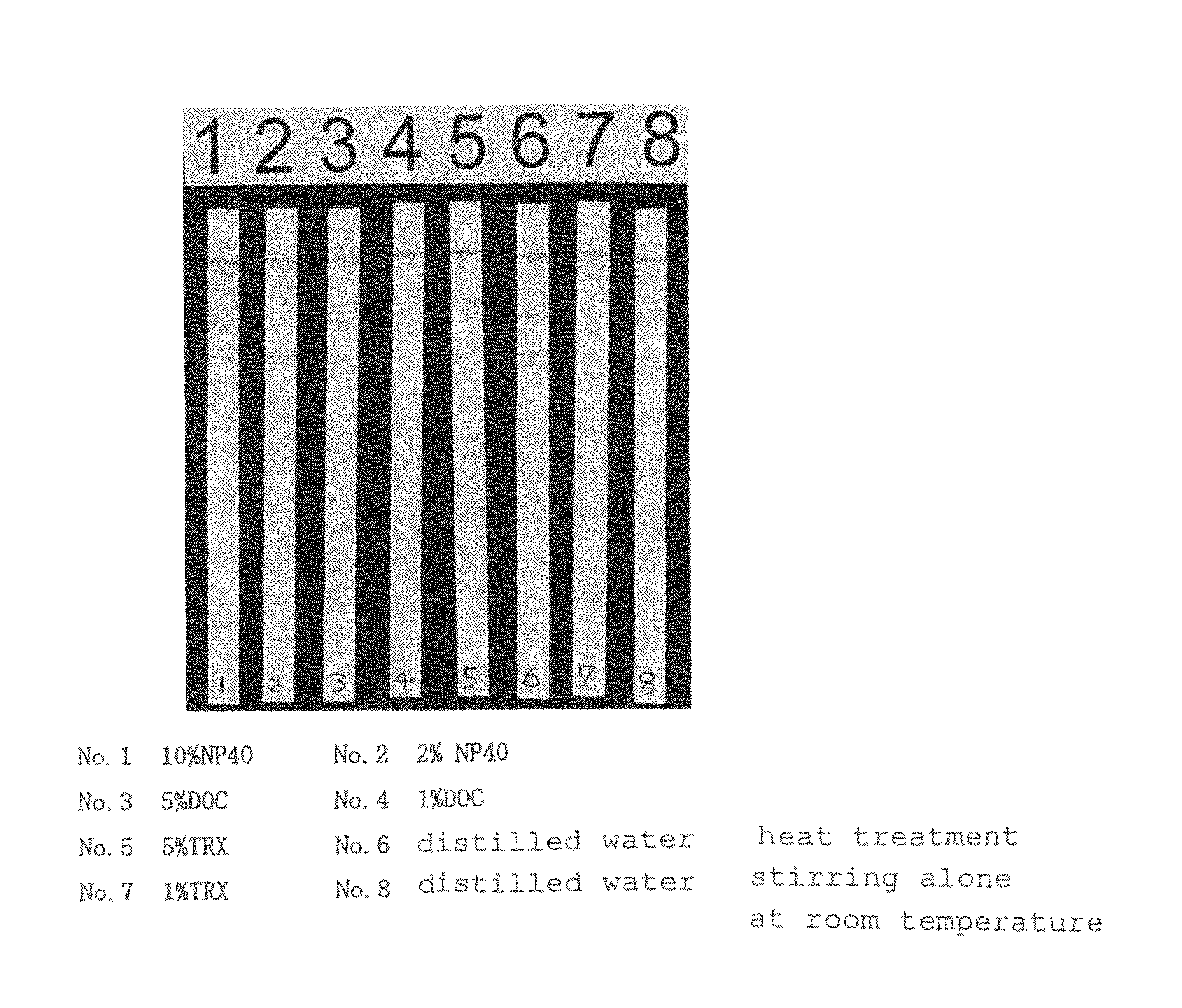
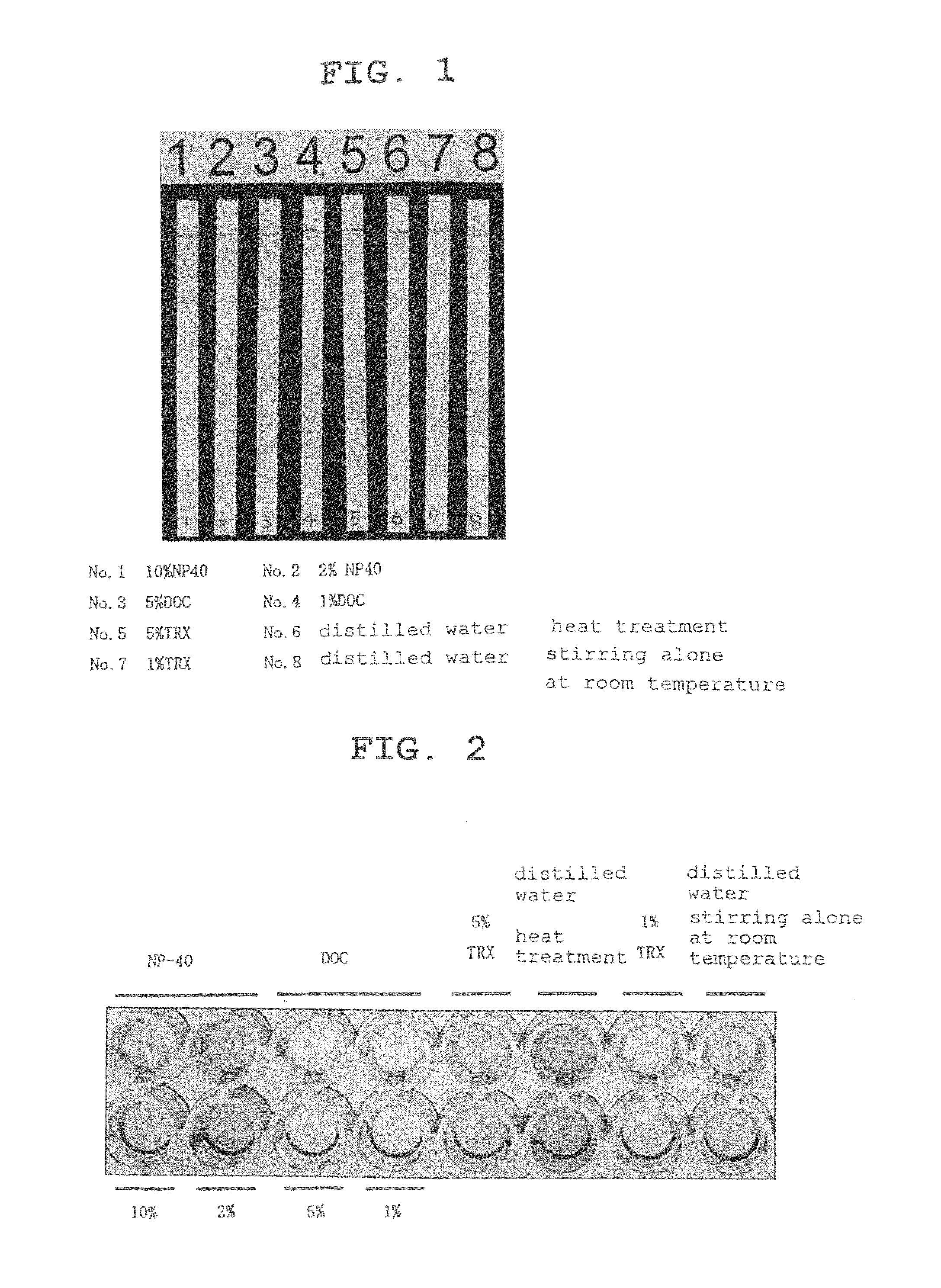
[0104]As the surfactants, sodium deoxycholate (hereinafter DOC), which is an anionic surfactant, and NP-40 (polyoxyethylene nonylphenyl ether) and TNX (polyoxyethylene iso-octylphenyl ether), which are non-ionic surfactants, were used.

[0105]5 wt % and 1 wt % aqueous solutions were prepared for DOC, 10 wt % and 2 wt % aqueous solutions were prepared for NP-40, and 5 wt % and 1 wt % aqueous solutions were prepared for TNX. A nail from a tinea unguium patient definitively diagnosed by the KOH method was divided into 8 almost equivalent amounts. Six out of 8 nails were each placed in an aqueous surfactant solution (300 μl), and the mixture was stirred at room temperature (about 20° C.) for 20 min. As one control, one nail was placed in 300 μl of distilled water, and the mixture was stirred at room temperature (about 20° C.) for 20 min. For the other control, one nail was placed in 300 μl of distilled water, and the mixture was subjected to a heat treatment by placin...

example 2

Detection of Tinea Fungi by Tinea Fungi Diagnosis Strip Test

[0106]From the samples after each treatment, 120 μl was applied to a test piece for a tinea fungi diagnosis strip test produced using the 0014 antibody. The specific method is as follows.

1) Production of Antibody Solid-phased Support

[0107]To produce an antibody solid-phased support on which the 0014 antibody has been linearly solid-phased, a nitrocellulose sheet (Millipore, HiFlow Plus) was cut into 5 mm×20 mm, and the 0014 antibody solution was applied to the sheet at 10 mm from the lower end with BioJet Q3000 (Biodot), and dried at room temperature for 2 hr.

2) Preparation of Colloidal Gold Particle-labeled Material Holding Carrier

a. Colloidal Gold Particle-labeled 0014 Antibody

[0108]To a dispersion (100 mL) of colloidal gold having a particle size of 40 nm, which was adjusted to pH6.0 with 0.1% K2CO3 solution was added a monoclonal antibody 0014 solution (400 μg), and the mixture was admixed at room temperature. Then the ...

example 3

Detection of Antigen by ELISA

[0112]A treatment liquid sample similar to that prepared in Example 1 was measured by ELISA. The method was as follows.[0113]1) solid phasing of antibody: The 0014 antibody was diluted with 50 mM carbonate buffer (pH 9.6) to a concentration of 20 μg / ml, dispensed to each well of a 96 well microplate (#9018, Corning Incorporated) by 50 μl, and incubated overnight at 4° C.[0114]2) blocking: An aqueous Yukijirushi BlockAce 25% solution was prepared, dispensed by 300 μl, and incubated at room temperature for 1 hr.[0115]3) washing: PBS containing 0.05% Tween 20 (Nacalai Tesque) was dispensed by 300 μl, and washed by repeating stirring and discarding 3 times.[0116]4) antigen-antibody reaction with sample: A sample was added to each well by 50 μl, and the mixture was incubate at room temperature for 1 hr.[0117]5) The washing operation similar to the above-mentioned 3) was performed 3 times.[0118]6) A biotinylated 0014 antibody was diluted to 1 μg / ml with 10% aq...


PUM

PropertyMeasurementUnit
temperatureaaaaaaaaaa
carbon numberaaaaaaaaaa

Abstract

The present invention provides a method of detecting dermatophyte, which does not require a complicated operation such as an enzyme treatment and a heat treatment.The present invention provides a method of detecting dermatophyte, including a step of extracting a dermatophyte component from a sample with a treatment liquid containing a non-ionic surfactant or a zwitterionic surfactant, and a kit for diagnosing dermatophyte infection, containing a treatment liquid comprising the above surfactant, and an antibody specifically recognizing a dermatophyte component, which are housed in separate containers.

Description

[0001]This application is a U.S. national stage of International Application No. PCT / JP2007 / 055567 filed Mar. 19, 2007.TECHNICAL FIELD[0002]The present invention relates to a non-heating detection method of dermatophyte, and to the diagnosis field of tinea.BACKGROUND ART[0003]Tinea, what is called athlete's foot, is dermatomycosis caused by infection of the skin with dermatophytes such as Tricophyton rubrum (hereinafter to be abbreviated as T. rubrum), Tricophyton mentagrophytes (hereinafter to be abbreviated as T. mentagrophytes), Microsporum canis (hereinafter to be abbreviated as M. canis), Epidermophyton floccosum (hereinafter to be abbreviated as E. floccosum) and the like. The main fungal infection site is interdigit of foot. However, genital, corpus and the like are also infected therewith, causing inflammation and intense pruritus in the infection site.[0004]The diagnosis of tinea is mainly based on potassium hydroxide (KOH) direct microscopic examination including dissolvin...

Claims


Application Information

Patent Timeline
Patent Type & Authority Patents(United States)
IPC IPC(8): G01N33/00G01N33/569G01N33/53C12Q1/68
CPCG01N33/56961G01N2333/37G01N2800/20
Inventor NORIKI, SAKON
Owner MARUHO
Why Eureka
Try Eureka