
Method for correcting pathological configurations of segments of the lower extremities and device for realizing same

a technology of pathological configuration and lower extremity, applied in the field of medicine, can solve the problems of partial loss of motor skills, hypotrophy of pelvic girdle and lower extremity muscles, and limited use of said method, and achieve the effect of correction only

Inactive Publication Date: 2015-08-11
ZAKRYTOE AKTSIONERNOE OBSCHESTVO NAUCHNO PROIZVODSTVENNY TSENTR OGONEK
View PDF13 Cites 14 Cited by
  • Summary
  • Abstract
  • Description
  • Claims
  • Application Information

AI Technical Summary

Benefits of technology

The modular exoskeleton device effectively corrects pathological configurations in children and adults, allowing for free movement and preserving active joint function, with the ability to adapt to changing body dimensions and address various combinations of pathological conditions.

Problems solved by technology

However, the utility of said method is limited and it can be used, as a rule, in children, primarily those of young age.
Moreover, utilization of said method assumes that the user will stay, for a long period, in a position that, throughout the correction process, precludes free movement, which leads to partial loss of motor skills and hypotrophy of muscles of the pelvic girdle and the lower extremities.
However, this correction can only be achieved after many years of physical exercise, and only to an insignificant extent.
Moreover, the utility of said method and device is limited inasmuch as they can be used in adults and older children, but cannot be used for correction of pathological configurations of segments of the lower extremities in younger children, including those suffering from infantile cerebral palsy (ICP).
Further, fixation of the user in said exercise machine limits free movements of the user, whereas holding the lower extremities in a given position precludes the development of motor skills and is not conducive to unassisted movement of the user.
However, said device comprises a complicated construction, and is custom-produced for a given user as the thigh and tibia sections have fixed lengths which precludes one—if necessary—from changing this device's linear dimension.
Thus, the utility of said device is limited.
Moreover, said device may only be used as a single construction.
In the event when it is not medically necessary to assist the movement of all regions of the lower extremities, utilization of such a device is not reasonable.
However, the utility of said method and device is limited as they can be used to move adults and older children, but cannot be used for correction of pathological configurations of segments of the lower extremities in younger children, including those suffering from infantile cerebral palsy, since the device is good only for individual use due to the fact that it is manufactured in accordance with the specific dimensions of a given user without the possibility of correcting the device while applying this method.
This precludes the possibility of using said device and method in children whose body dimensions change substantially within a short time period.
Moreover, use of said device leads to weakening and hypotrophy of muscles of the lower extremities.
The utility of the known device is limited and it cannot be used by individuals with hip dislocations and subluxations because the pelvic belt of the known device only allows positioning of the device on the user's body, and does not allow centering the femoral head in the hip socket.
The lack of ability to center the femoral head, in its turn, does not allow applying an adequate load onto the entire surface of the user's lower extremity.
Using the known device, it is not possible to carry out correction of the rotation axis of the knee joint because the flat knee hinge used therein does not allow correction of the tibia's position relative to the thigh when the position of the tibia is abnormal.
Moreover, the known device does not allow adaptation to the user's anatomical characteristics because it is not possible to change the configuration of the flat connecting rods that takes into account, for example, the shape of the tibia or the thigh.
Further, said device features a complex construction, including hinge connections containing moving and rotating parts that are in direct proximity to the user's body, which practically precludes the use of this device in children, including younger children.

Method used


Image

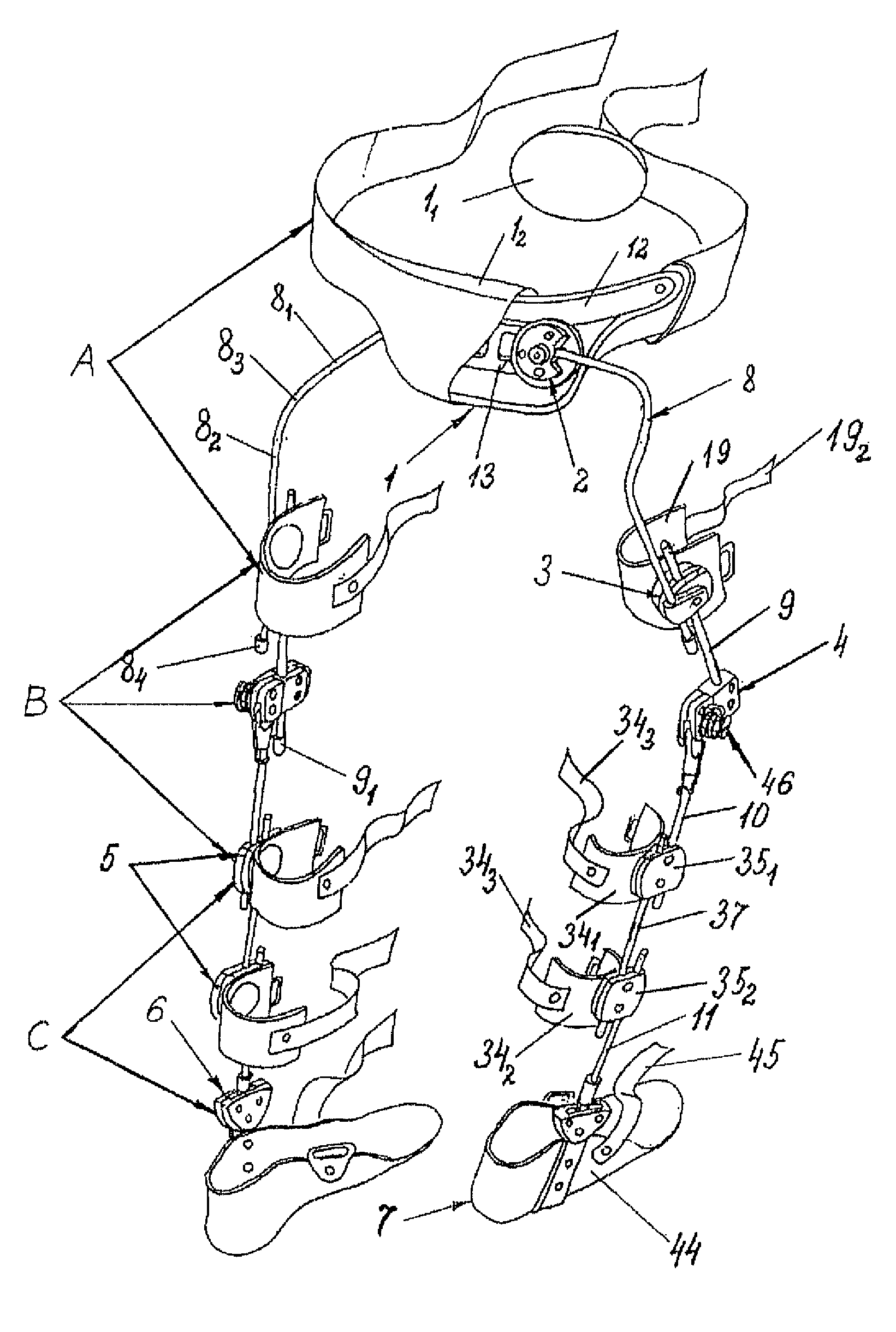
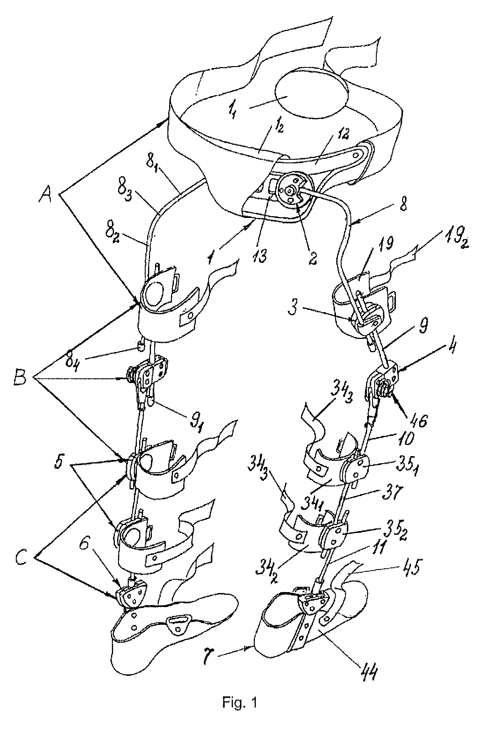
  • Method for correcting pathological configurations of segments of the lower extremities and device for realizing same
  • Method for correcting pathological configurations of segments of the lower extremities and device for realizing same
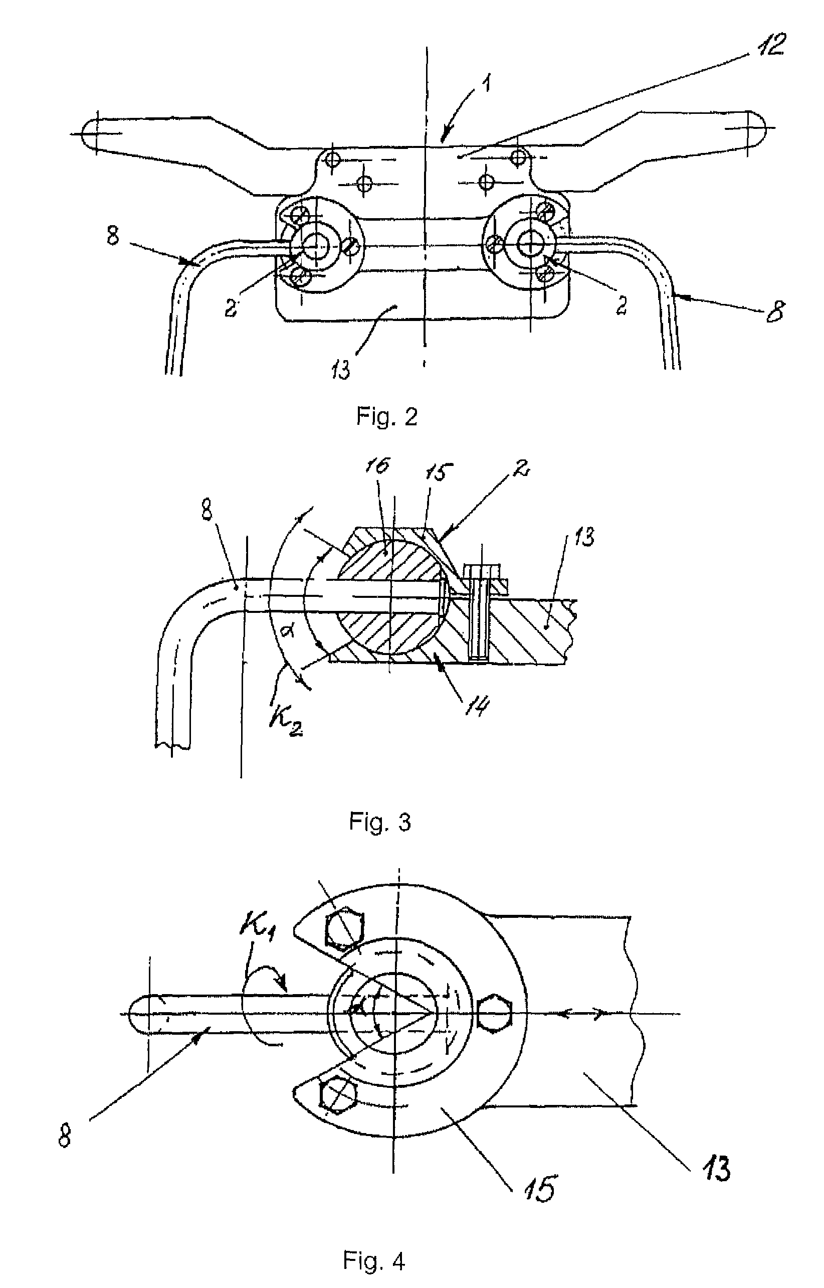
  • Method for correcting pathological configurations of segments of the lower extremities and device for realizing same

Examples


example 1

[0333]User N. R., age: 8.

[0334]Diagnosis: infantile cerebral palsy (thereafter ICP), spastic diplegia predominantly affecting the right extremities, severe form, adductor-prone positioning of the thighs, pathological flexion and valgus positioning of the tibias, more on the right, abductor-equine-planovalgus positioning of the feet.

[0335]On examination: the user does not stand or walk without assistance. Patient walks while supported on one arm or while leaning on two Canadian crutches in the position of triple bending with inner rotation of the thighs and leaning on front-inner regions of the feet. During walking, there is non-permanent crossing of lower extremities at the level of the lower third of the thigh. There is pronounced valgus positioning of the right tibia in the position “standing” and during locomotion; valgus positioning of the left tibia is less pronounced. In the position “standing” and during locomotion, there is notable pathological abduction of the feet, more so...

example 2

[0341]User K. K., age: 24.

[0342]Diagnosis: ICP, spastic diplegia of moderate severity predominantly affecting left extremities, secondary multi-dystrophic syndrome, adductor-prone positioning of the thighs, inner rotation of both lower extremities, valgus positioning of the left foot, pronounced equine-planovalgus positioning of the left foot.

[0343]On examination: the user walks without assistance yet with a pronounced rocking of the torso during walking, rotates the lower extremities inward with support on the entire right foot and front-inner region of the left foot. The thighs are adducted. The support phases during locomotion are not differentiated (there is no “rollover” from the heel part of the foot to the distal part of the foot during walking). In the standing position and during locomotion, moderate valgus positioning of the left tibia is evident. The right lower extremity is more supportive, the single-support period of the left lower extremity is shortened. There is nota...

example 3

[0349]User: A. A., age: 3.

[0350]Diagnosis: consequences of a closed craniocerebral injury, brain contusion, left-sided dissociative hemiparesis predominantly affecting the left lower extremity, abnormalities of posture in the frontal plane, flexion positioning of the left forearm, adductor-prone positioning of the left thigh, recurvation of the left tibia, adductor-equine-varus positioning of the left foot with adduction of the front region, anatomical shortening of the left lower extremity by 7 mm.

[0351]On examination: the user stands and walks without assistance. The walk is hemiparetic, non-elastic, with frontal rocking of the torso; during walking, leans on frontal-outer regions of the left foot, the phases of the left foot's support period are not differentiated. During walking, there is recurvation of the left tibia in the knee joint. Reciprocal movements of upper extremities are absent during walking. Active dorsiflexion of the left foot is absent.

[0352]The proposed exoskelet...


PUM


Abstract

The invention relates to a method for the step-by-step correction of the position of the longitudinal axes of segments of the lower extremities in the horizontal and / or frontal and / or sagittal planes, that comprises the simultaneous dosed correction of the imbalance of forces in the muscles supporting the corresponding joints, the reduction of the pathological configurations of the segments, and the fixation of corrected segments using at least one module of a modular exoskeleton capable of individual adjustment for bringing, during each step, the position of the longitudinal axis of each segment closer to the physiologically correct position, and for permitting movements about the corrected rotation axes of the joints within a predetermined range defined by the modular exoskeleton. The proposed device for correcting pathological configurations of segments of the lower extremities comprises a modular exoskeleton, the modules of which can be used independently or in various combinations. The modular exoskeleton comprises at least one adjustable pelvis belt (1, 47), at least one means (3, 51) for holding the thigh in a given position, at least one means (4, 89) for rotating the tibia, at least one means (5, 121) for holding the tibia in a given position, at least one means (6, 130) for correcting the position of the foot and the movement range thereof at the foot-tibia joint, at least one means (7, 146) for accommodating the foot, and a set of fixation members each in the form of a cylindrical rod (8, 9, 10, 11, 37, 52, 83, 90, 124, 141, 142) capable of linear positioning movement and / or rotation and / or free linear movement.

Description

CROSS REFERENCE TO RELATED APPLICATIONS[0001]This application is the National Stage of PCT / RU2010 / 000746 filed on Dec. 10, 2010, which claims priority under 35 U.S.C. § 119 of Eurasian Patent Organization No. 201000171 filed on Dec. 15, 2009, Eurasian Patent Organization No. 201001788 filed on Nov. 12, 2010, and Eurasian Patent Organization No. 201100041, filed on Nov. 26, 2010, the disclosures of which are incorporated by references. The international application under PCT article 21 (2) was not published in English.FIELD OF THE INVENTION[0002]The present invention is in the field of medicine, specifically traumatology, orthopedics, and neurology, and more specifically—methods for correcting pathological configurations of segments of the lower extremities and devices for realizing same.[0003]At the present time, due to worsening of the ecological environment, the impact of adverse factors on the population—including pregnant women—is rising and, as a consequence, propensity of the ...

Claims


Application Information

Patent Timeline
Patent Type & Authority Patents(United States)
IPC IPC(8): A61F5/00A61F5/01
CPCA61F5/0102A61F5/0123A61F5/0127A61F5/0193A61F5/02A61F5/05
Inventor CHUGUNOV, VITALY VIKTOROVICH
Owner ZAKRYTOE AKTSIONERNOE OBSCHESTVO NAUCHNO PROIZVODSTVENNY TSENTR OGONEK
Learn More