Methods of analyzing an H and E stained biological sample

a biological sample and h and e technology, applied in the field of methods of analyzing an h and e stained biological sample, can solve the problems of limited sample amount, difficulty in performing various analyses required for complete characterization of disease, and limited ability to determine relative characteristics of targets

Active Publication Date: 2015-11-03
LEICA MICROSYSTEMS CMS GMBH
View PDF36 Cites 4 Cited by
  • Summary
  • Abstract
  • Description
  • Claims
  • Application Information

AI Technical Summary

Problems solved by technology

Additionally with early cancer detection sample size is diminishing, making it difficult to perform various analyses required for complete characterization of disease.
While valuable, many of these current techniques may detect only a few targets at one time (such as IHC or fluorescence-based Western blots where number of targets detectable is limited by the fluorescence-based detection system) in a single sample.
Further analysis of targets may require use of additional biological samples from the source, limiting the ability to determine relative characteristics of the targets such as the presence, absence, concentration, and / or the spatial distribution of multiple biological targets in the biological sample.
Moreover, in certain instances, a limited amount of sample may be available for analysis or the individual sample may require further analysis.
A major problem with using the previously H&E stained slide for IHC and FISH is the interference from H&E stains in both chromogenic detection in IHC and fluorescence detection for IF and FISH.
While this has allowed IHC with chromogen detection, IF and FISH are not feasible due to strong residual fluorescence from eosin.
Removal of residual eosin fluorescence has not been feasible with many different techniques attempted to date.

Method used


Image

  • Methods of analyzing an H and E stained biological sample
  • Methods of analyzing an H and E stained biological sample
  • Methods of analyzing an H and E stained biological sample

Examples


example 1

Preparation of Tissue Samples

[0114]Human breast tissue array samples were obtained as tissue slides embedded in paraffin from Clarient, Huntsville, Ala. Slides were baked at 60° C. for 15 minutes and then H&E stained.

example 2

H&E Staining

[0115]All the slides were stained with hematoxylin and eosin following the standard regressive protocol listed below. After staining, images were obtained as described below in Table 1. In the procedure the slide with tissue is baked at 60° C. for 15 minutes prior to the first XYLENE step. The Hematoxylin and Eosin were filtered before each use. Coverslip was applied directly out of xylene to prevent the slide from drying before coverslipping.

[0116]

TABLE 1Standard Regressive protocol H&E StainingSolutionTimeVendorManufacture / Catalog #Xylene5minutesFisherFisher Chemical #C8H10Sci.Xylene3minutesFisherFisher Chemical #C8H10Sci.100% Alcohol1minuteVWREMD-HARLECO, 100% Alcohol blend #34172-020100% Alcohol1minuteVWREMD-HARLECO, 100% Alcohol blend #34172-02195% Alcohol1minuteVWREMD-HARLECO, Dehydration Alcohol 95%, #6534895% Alcohol1minuteVWREMD-HARLECO, Dehydration Alcohol 95%, #6534980% Alcohol1minuteVWREMD-HARLECO, Dehydration Alcohol 95%, #65350DI Water20-30secondsHEMATOXYLI...

example 3

Imaging

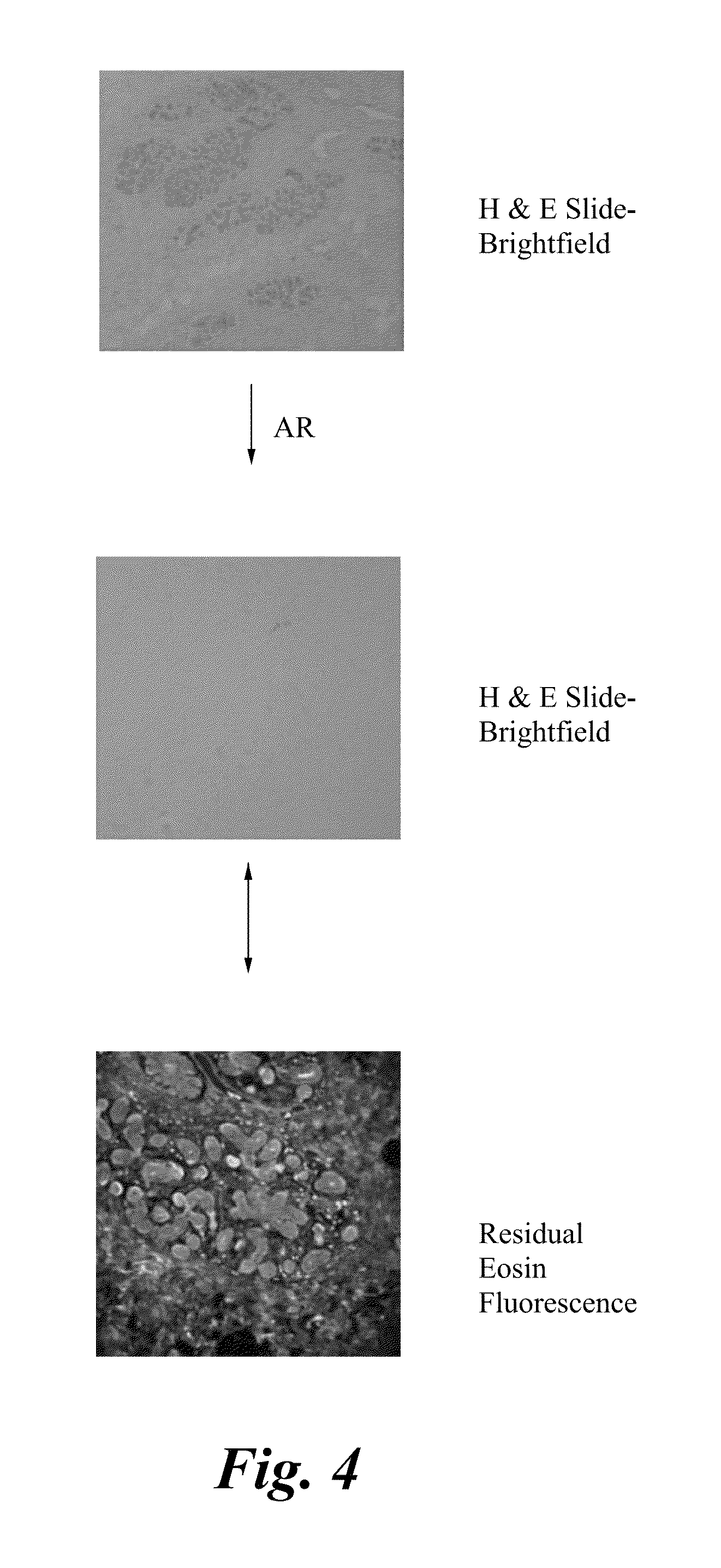
[0117]Brightfield and Fluorescence images were taken for all the slides using Leica brightfield microscope and Olympus microscope respectively. For fluorescence, eosin stained images were collected in Cy2 (Ex: 482 / 35, Em: 536 / 40), Cy3 (Ex: 531 / 40, Em: 593 / 40), and Cy5 (Ex: 628 / 40, Em: 692 / 40) channels.


PUM

PropertyMeasurementUnit
wavelengthaaaaaaaaaa
temperatureaaaaaaaaaa
wavelengthsaaaaaaaaaa

Abstract

Methods comprising the use probing multiple targets in a H&E stained biological sample are provided. The methods include the steps of providing a hematoxylin and eosin stained biological sample containing multiple targets, observing the sample, removing the hematoxylin and partially removing the eosin by washing the sample, contacting the sample with a borate salt, and irradiating the sample to remove the residual eosin fluorescence. The method further includes the optionally performing the additional steps of binding at least one probe to one or more targets to the sample, observing a signal from the probe and contacting the sample with a bleaching agent. The process of binding, observing and bleaching may be iteratively repeated.

Description

CROSS-REFERENCE TO RELATED APPLICATIONS[0001]This application is a continuation in part of U.S. patent application Ser. No. 13 / 336,409 filed on Dec. 23, 2011 and is herein incorporated by reference.BACKGROUND[0002]Traditionally, hematoxylin and eosin staining (H&E) is one of the most common staining methods in histology. The morphology from the H&E is most widely used in medical diagnosis for cancer detection. However, increasingly molecular analysis of tissue by immunohistochemistry (IHC) or immunofluorescence (IF) and fluorescence in situ hybridization have become an essential part of cancer diagnosis in addition to morphological assessment. With great strides in targeted therapies, detailed molecular assessment of cancer tissue is fast becoming a requirement. Additionally with early cancer detection sample size is diminishing, making it difficult to perform various analyses required for complete characterization of disease.[0003]While valuable, many of these current techniques ma...

Claims


Application Information

Patent Timeline
Patent Type & Authority Patents(United States)
IPC IPC(8): G01N33/53G01N33/532G01N33/58G01N21/76G01N1/30G01N33/533G01N21/64
CPCG01N1/30G01N33/582G01N21/64G01N21/6428
Inventor SOOD, ANUPKENNY, KEVIN BERNARDNATARAJAN, ARUNKUMARKAANUMALLE, LAKSHMI SIREESHA
Owner LEICA MICROSYSTEMS CMS GMBH
Why Eureka
Try Eureka