Peripheral neural interface via nerve regeneration to distal tissues

a neural interface and nerve technology, applied in the field of peripheral neural interface via nerve regeneration to distal tissues, can solve the problems of poor utilization of powered prostheses, unoptimized elimination of native tissue functionality for the greater good, and developers still struggle with issues, so as to maximize the potential for natural neural control and enhance the prosthetic control

Active Publication Date: 2016-10-25
MASSACHUSETTS INST OF TECH
View PDF22 Cites 3 Cited by
  • Summary
  • Abstract
  • Description
  • Claims
  • Application Information

AI Technical Summary

Benefits of technology

[0009]In another embodiment, the method of reversing impairment of a human limb includes transecting a nerve associated with the impairment of the limb of an individual to thereby form proximal and distal ends of the transected nerve. The proximal and distal ends of the transected nerve are placed into proximal and distal ends of a microchannel array, thereby causing the nerve to regenerate through the microchannel array. Sensory afferent information of the regenerated nerve is recorded using sensing electrodes within a plurality of afferent microchannels of the microchannel array. Motor efferent information is stimulated to provide efferent motor stimulus to the nerve using stimulating electrodes within the plurality of efferent microchannels of the microchannel array. The stimulating electrodes are electrically connected to a motor controller of a device. The sensing electrodes are electrically connected a sensory controller of the device, wherein the sensor controller is linked to at least one sensor of the biological limb that detects application of at least one of position, velocity, acceleration, and force of the biological limb, and whereby the sensory controller transmits detection of the position, velocity, acceleration, and force of the biological limb to the motor controller, and whereby the motor controller applies electrical stimulation via the stimulating electrodes, thereby reversing impairments of the human limb.
[0011]In still another embodiment, the method of the invention includes reversing the impairment of an amputated limb, including inserting a distal end of at least one transected nerve of an amputated limb into a proximal end of a microchannel array, placing at least one member of the group consisting of skin and muscle end organ at the distal end of the microchannel array, thereby causing the nerve to regenerate through the microchannel array and to innervate the at least one end organ. Efferent motor information of the regenerated nerve is recorded using sensing electrodes within a plurality of afferent microchannels of the microchannel array. The regenerated nerve is stimulated with afferent sensory information using stimulating electrodes within a plurality of afferent microchannels of the microchannel array. The sensing electrodes are electrically connected to a motor controller of a device. The stimulating electrodes are electrically connected to a sensory controller of the device, wherein the motor controller is linked to at least one sensor of the device that detects application of at least one member selected from the group consisting of position, velocity, acceleration, and force of the device, and whereby the motor controller transmits detection of the position, velocity, acceleration, and force by applying electrical stimulation via the stimulating electrodes, thereby providing the individual with a sensation simulating sensory feedback from the device, and reversing impairment of the amputated limb.
[0015]The surgical reconstruction methodology and implantable system of the invention significantly increases the potential for natural neural control of prostheses, such as artificial limbs and functional electrical stimulation devices. The system utilizes the neural activity within the residual biological limb generated in the peripheral nerves and / or the electromyographic activity generated through muscle tissue activation. Such nerve and muscle tissues may be native to the residual limb, or they may be relocated to the residual limb through a plurality of surgical manipulations. Such manipulations may include free muscle grafts or pedicle muscle grafts, which may include intact attached nerves and / or vasculature. Additionally, the musculature could be derived from transplanted muscle precursor cells or cultured muscle tissue. For amputation limb patients, the system includes means to record one or multiple independent channels of neural motor activity that can control the various degrees of freedom present in advanced powered prosthetic limbs. Further, the method of the invention provides for the possibility of sensory information input from a controlled external prosthesis back into the nervous system. With regard to cutaneous sensory feedback, this can be by electrically activating sensory nerves through a nerve cuff and / or microchannel array directly, or by applying mechanical stimulation to the native skin of the residual limb or other cutaneous tissue, such as fingertip skin, that has been relocated by a grafting procedure to the residual limb.
[0016]The invention also includes means to provide proprioceptive feedback to the amputee. This can be achieved through direct electrical activation of muscle and tendon afferent nerve fibers using the microchannel array. The invention also allows relocated antagonistic muscle pairs to mechanically interact with each other in a reciprocal push-pull fashion, just as would occur if the agonist / antagonist muscle pair were attached to the opposing sides of a joint. This approach allows the muscle proprioceptive endings that are intrinsic to those muscles to be activated by a normal stretch stimulus that occurs with intact muscles operating around the same joint.

Problems solved by technology

Despite these engineering accomplishments, developers still struggle with the issue of how to provide the prosthesis user with methods for coordinating the simultaneous control of all of the joints that are involved with, for example, object manipulation in the upper extremity case, or standing and walking in the lower extremity case.
The resulting motion for most activities is thus awkward, time consuming, and tedious, since it breaks up any compound arm and hand movement into serial positioning steps, resulting in poor utilization of powered prostheses.
However, commercial lower-extremity prostheses typically do not utilize EMG as a source of control signals.
Having to eliminate the functionality of any native tissue for the greater good is not optimal.
There are also some limitations regarding how far away a given nerve can be moved in order to connect it to a suitable muscle target.
Finally, the use of surface recorded EMG and contiguous muscle targets can lead to inconsistent signal amplitudes and objectionable channel crosstalk (T. A. Kuiken, M. M. Lowery, and N. S. Stoykov, “The effect of subcutaneous fat on myoelectric signal amplitude and cross-talk,” Prosthetics and Orthotics International, vol.
Over time, however, it is still necessary to “re-tune” the system, which is a substantial inconvenience.

Method used


Image

  • Peripheral neural interface via nerve regeneration to distal tissues
  • Peripheral neural interface via nerve regeneration to distal tissues
  • Peripheral neural interface via nerve regeneration to distal tissues

Examples


first embodiment

Description of Specific Embodiments of the Invention

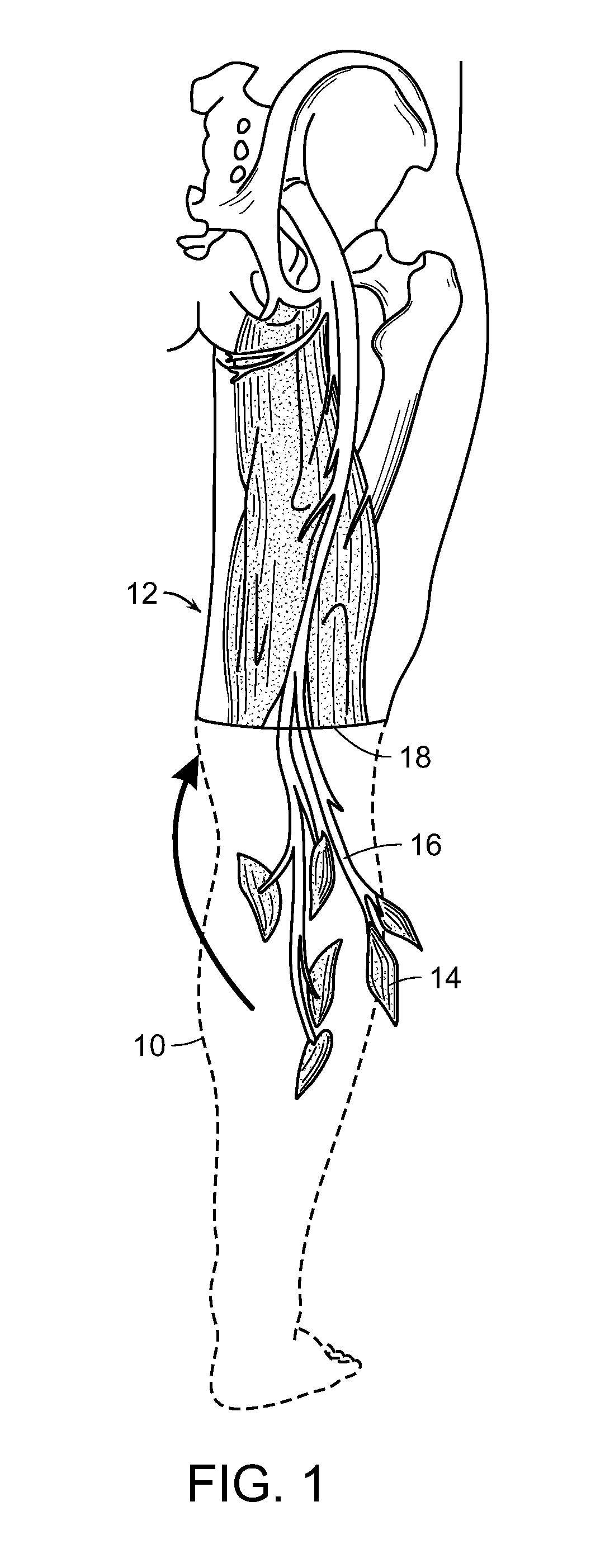
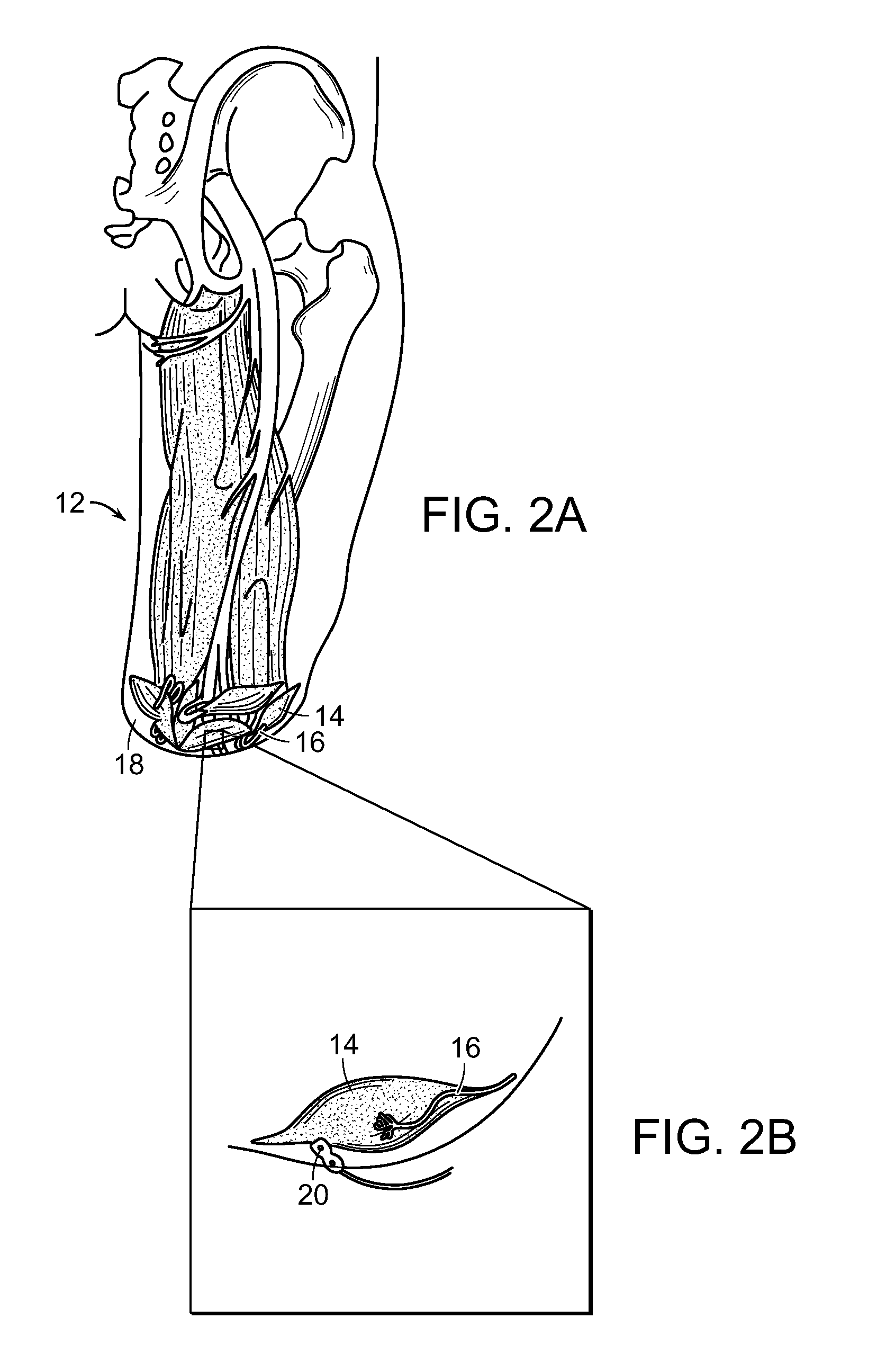
[0069]FIGS. 1, 2A and 2B are schematic representations of one embodiment of the method of the invention that includes the method of the restoring at least partial function of a human limb. At least a portion 10 (shown in outline) of an injured or diseased human limb 12 from an individual is surgically removed, leaving intact at least a portion of selected muscles 14 from the damaged portion of the human limb, including blood vessels and nerves 16 associated with that portion of the selected muscles 14. The selected muscles are transplanted to a surgical site 18 of the individual where the human limb was removed. The transplanted selected muscles 14 and associated nerves 16 are then contacted with electrodes 20 (FIGS. 2A and 2B), whereby signals can be transmitted to and from nerves 16 at the transplanted muscles 14 to thereby control a prosthetic limb (not shown) that is linked to the electrodes and extends from the surgical site 1...

second embodiment

[0088]FIGS. 4-6D are schematic representations of a second embodiment of a method of the invention for restoring at least partial function of a human limb. Specifically, FIG. 4 is a schematic representation of surgical removal of a portion 22 (in outline) of an injured or diseased human limb 24, in outline, leaving intact a portion of selected muscles 26 and selected glabrous skin patches 28, including blood vessels and nerves 30 associated with those portions of the selected muscles 26 and glabrous skin patches 28, according to another embodiment of the invention. Alternatively, or in addition, the nerve can be a new regernative innervation nerve. “New regenerative innervation nerve,” as that term is employed herein, is a nerve that has regenerated or grown into muscle or skin cells. Similarly, an “end organ,” as that term is employed herein, is a tissue body into which a nerve regenerates. Thus, a muscle body or a skin body into which a nerve has regenerated are end organs. FIG. 5...

third embodiment

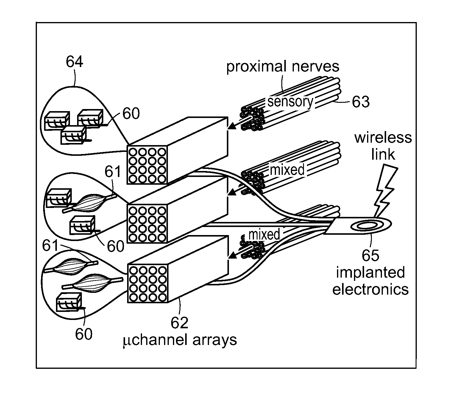
[0094]A third embodiment of the method of restoring at least partial function of a human limb of the invention includes sensory feedback and, as illustrated in FIGS. 7A-7B, the use of implanted electrodes 46 at muscles 48 and other implanted electronics 55 for registering EMG activity. FIG. 7A is a schematic representation of contacting native sensory innervation 50 of a skin patch 52 with nerve cuffs 54, wherein the nerve cuffs 54 are linked to a controller 56, and selectively stimulates native sensory innervation by actuating the nerve cuffs 54 with the controller 56. Nerve cuffs 54 are linked to implanted electronics 55 which are, in turn connected, such as by a wireless connection, to controller 56, as shown in FIG. 7A. FIG. 7B is another schematic representation of the embodiment of the invention shown in FIG. 7A, showing placement of implanted electronics and the nerve cuffs of FIG. 7A. In this embodiment, relocated distal skin 52 and its native sensory nerves 50 are packaged ...


PUM


Abstract

At least partial function of a human limb is restored by surgically removing at least a portion of an injured or diseased human limb from a surgical site of an individual and transplanting a selected muscle into the remaining biological body of the individual, followed by contacting the transplanted selected muscle, or an associated nerve, with an electrode, to thereby control a device, such as a prosthetic limb, linked to the electrode. Simulating proprioceptive sensory feedback from a device includes mechanically linking at least one pair of agonist and antagonist muscles, wherein a nerve innervates each muscle, and supporting each pair with a support, whereby contraction of the agonist muscle of each pair will cause extension of the paired antagonist muscle. An electrode is implanted in a muscle of each pair and electrically connected to a motor controller of the device, thereby simulating proprioceptive sensory feedback from the device.

Description

RELATED APPLICATION(S)[0001]This application claims the benefit of U.S. Provisional Application No. 61 / 894,040, filed on Oct. 22, 2013 and U.S. Provisional Application No. 62 / 019,266, filed on Jun. 30, 2014. The entire teachings of the above application(s) are incorporated herein by reference.BACKGROUND OF THE INVENTION[0002]Recent advances in prosthetic limbs include the provision of multiple degrees of freedom as well as powered actuators that have the potential to provide substantially greater functionality than the passive devices that existed just a decade ago. Despite these engineering accomplishments, developers still struggle with the issue of how to provide the prosthesis user with methods for coordinating the simultaneous control of all of the joints that are involved with, for example, object manipulation in the upper extremity case, or standing and walking in the lower extremity case. This deficit was first apparent for upper extremity prostheses, which now can provide e...

Claims


Application Information

Patent Timeline
Patent Type & Authority Patents(United States)
IPC IPC(8): A61N1/00A61F2/72A61B5/04A61N1/05A61F2/68A61N1/36A61B5/0488A61F2/60
CPCA61F2/72A61F2/68A61N1/0551A61N1/36003A61B5/04888A61F2002/607A61F2002/6827A61B5/0028A61B5/0031A61B5/40A61B5/4029A61B5/4041A61B5/4047A61B5/4052A61B5/441A61B5/45A61B5/48A61B5/4824A61B5/4827A61B5/4851A61B5/68A61B5/6801A61B5/6846A61B17/1128A61B2017/1135A61F2002/6872A61B5/24A61B5/389A61F2/482
Inventor HERR, HUGH M.RISO, RONALD R.SONG, KATHERINE W.CASLER, JR., RICHARD J.CARTY, MATTHEW J.
Owner MASSACHUSETTS INST OF TECH
Why Eureka
Try Eureka