Magnetic resonance examination system with preferred settings based on data mining

a magnetic resonance imaging and data mining technology, applied in the direction of reradiation, measurement using nmr, instruments, etc., can solve the problems of inferior image quality below the requested quality standards, difficult and tedious scan parameters selection, and inability to use advanced imaging techniques

Active Publication Date: 2017-02-14
KONINK PHILIPS ELECTRONICS NV
View PDF22 Cites 4 Cited by
  • Summary
  • Abstract
  • Description
  • Claims
  • Application Information

AI Technical Summary

Benefits of technology

[0017]As a consequence, any MR imaging protocol received via the user interface can be categorized into a certain MR scan type which in turn permits to provide an elegant possibility to provide statistical information with respect to this MR scan type, wherein the statistical information was obtained by analysis of previous MR scans.
[0020]In accordance with a further embodiment of the invention, the statistical information comprises statistics on ranges of modifications of individual scan parameters of the scan type. For example, the statistics on ranges of modifications of individual scan parameters of the MR scan type further comprise threshold ranges for said scan parameters. Thus, based on previously performed MR imaging scans, typical ranges of scan parameters are identified, such that a user may be assisted in selecting appropriate parameter values when adapting his imaging sequence by providing these ranges. Consequently, this prevents the user to accidentally enter unusual scan parameters which are completely ‘out of range’.
[0022]Additionally or alternatively it may be possible to provide for example the threshold ranges to the user interface which has the advantage that the user is assisted in choosing the most appropriate parameters since he will intuitively select the parameters to be within the threshold range.

Problems solved by technology

As a consequence, in these conditions the optimal choice of scan parameters is a difficult, tedious and often iterative task, even for expert users.
In other cases, the appropriate choice of scan parameters results in inferior image quality below the requested quality standards.
Another consequence is that advanced imaging techniques are not used as often as they could be, because the technician may not be aware of the suitable techniques to solve a particular image quality problem or to address a particular patient imaging need.
Consequently, this method permits to make imaging protocols available, however with the drawback that only ‘prefabricated’ protocols are provided, such that with respect to the individual circumstances with respect to an imaging procedure a user is still required to adapt the scan parameters of the selected MR scan protocol in an individual manner.
Consequently, the optimal choice of scan parameters is still difficult even for expert users.

Method used


Image

  • Magnetic resonance examination system with preferred settings based on data mining
  • Magnetic resonance examination system with preferred settings based on data mining
  • Magnetic resonance examination system with preferred settings based on data mining

Examples


Embodiment Construction

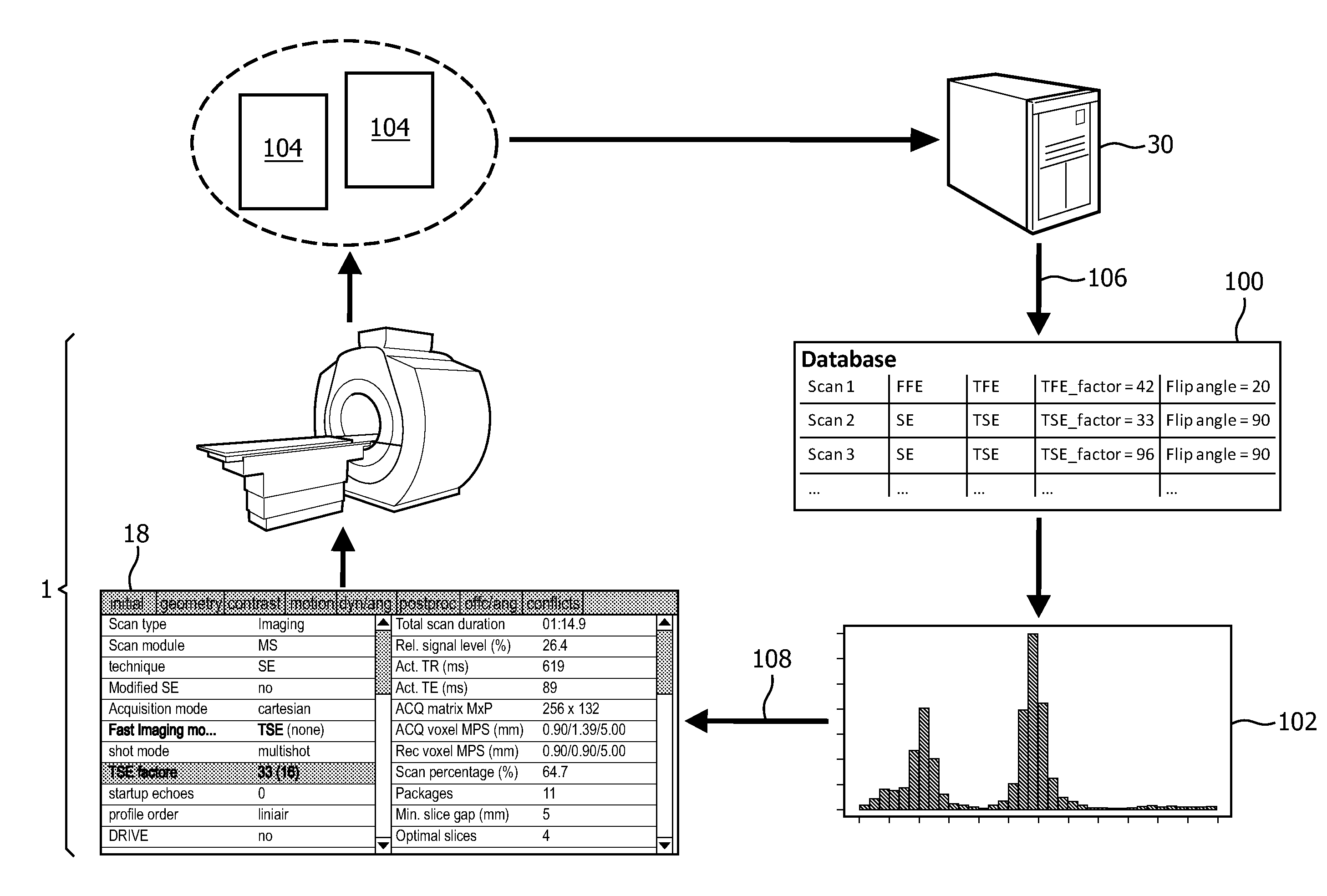
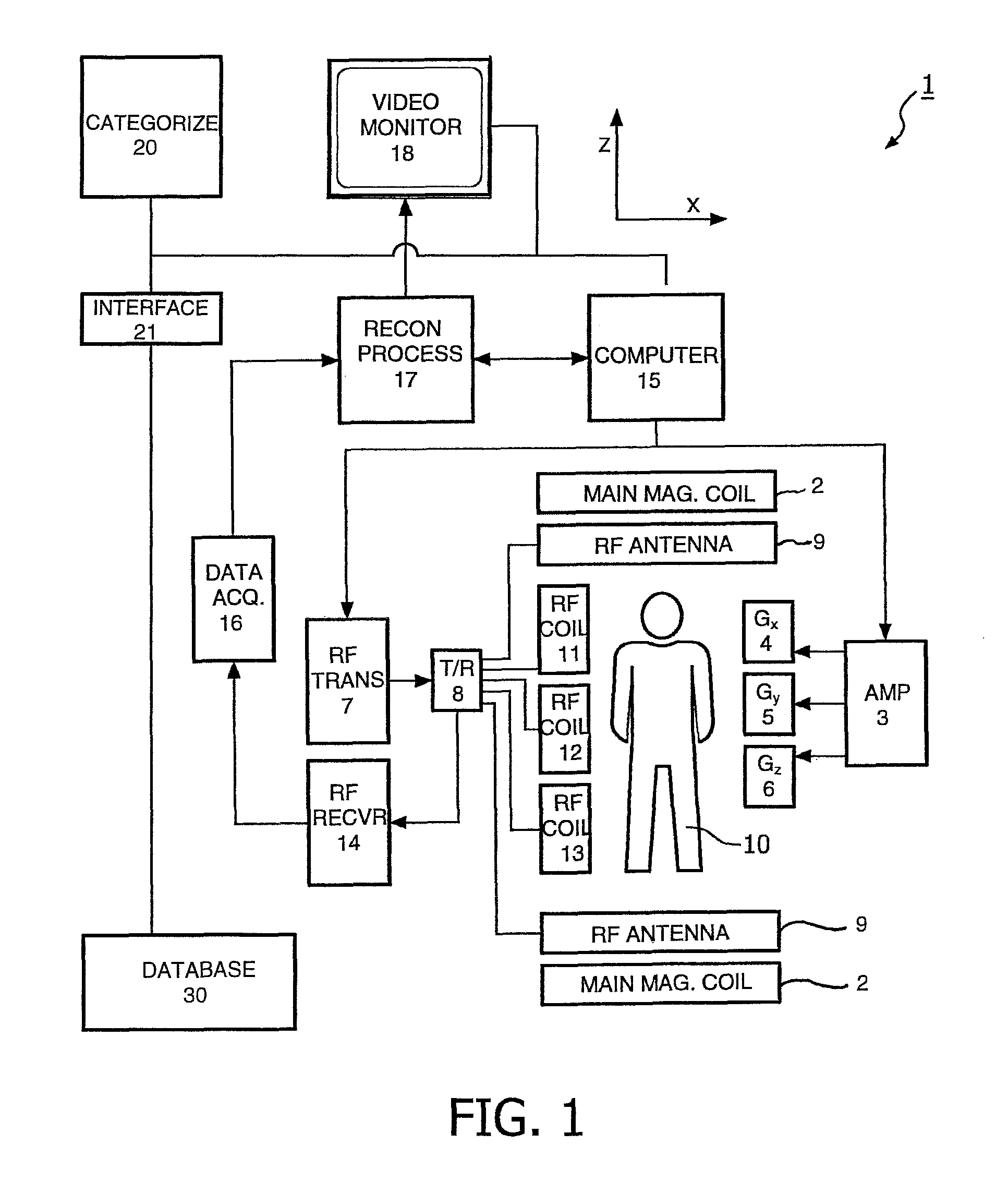
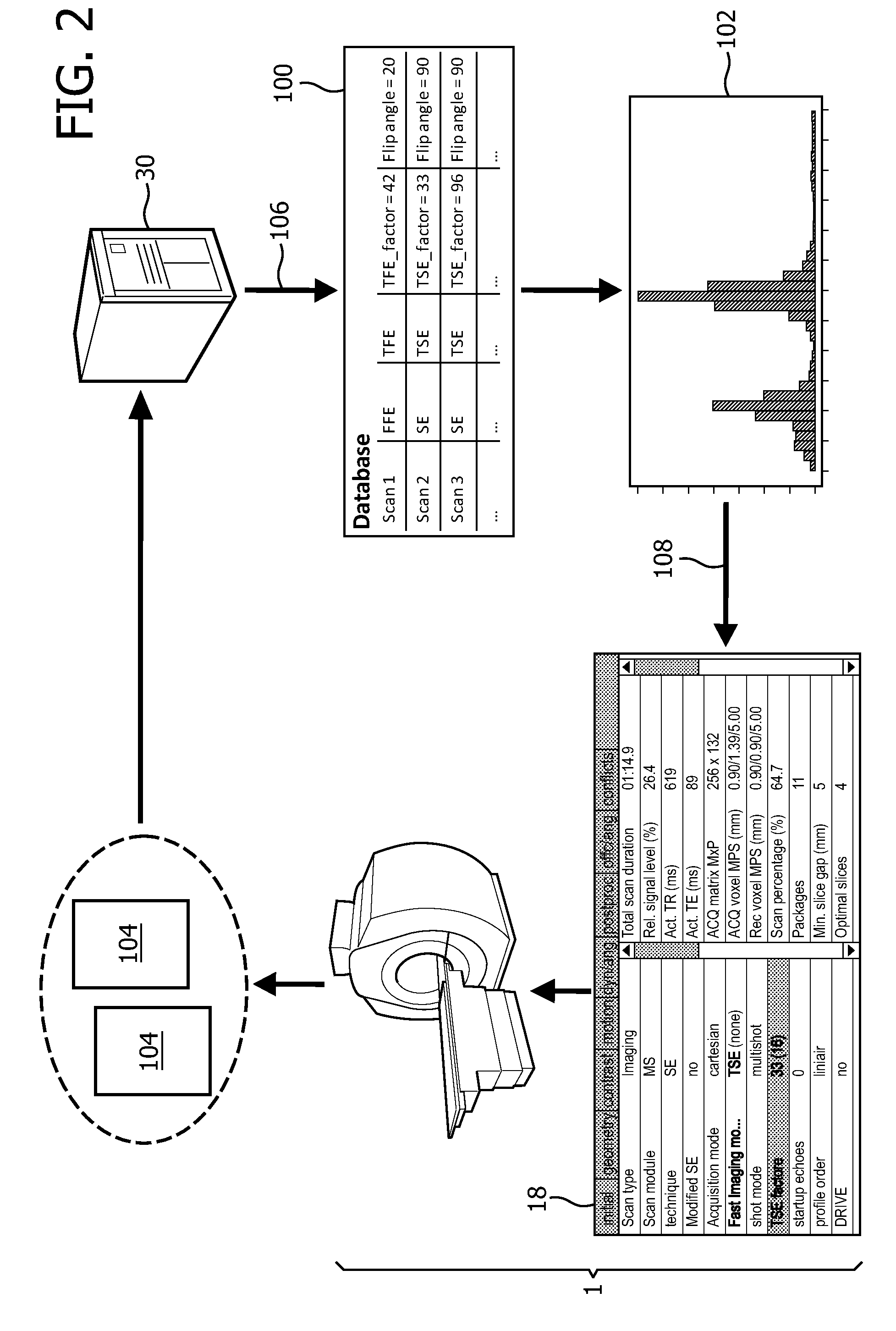
[0044]With reference to FIG. 1, an MR imaging system 1 is shown. The system comprises superconducting or resistive main magnet coils 2 such that a substantially uniform, temporarily constant main magnetic field BO is created along a z-axis through an examination volume.

[0045]The magnetic resonance system applies a series of RF pulses and switched magnetic field gradients to invert or excite nuclear magnetic spins, induce magnetic resonance, refocus magnetic resonance, manipulate magnetic resonance, spatially or otherwise encode the magnetic resonance, saturate spins and the like to perform MR imaging.

[0046]More specifically, a gradient pulse amplifier 3 applies current pulses to selected ones of whole body gradient coils 4, 5 and 6 along x, y and z-axes of the examination volume. An RF transmitter 7 transmits RF pulses or pulse packets, via a send / receive switch 8 to an RF antenna 9 to transmit RF pulses into the examination volume. A typical MR imaging sequence is composed of a pac...


PUM


Abstract

A magnetic resonance imaging scan using a MR scanner receives via a user interface a MR imaging protocol categorizable into a MR scan type of a predefined set of MR scan types. Further, a database is queried by providing to the database scan information permitting the database to identify the MR scan type of the MR imaging protocol. Statistical information on the MR scan type which can include statistics on modifications of individual scan parameters of the MR scan type is received from a database, and the statistical information is provided to the user interface. Modifications of the MR imaging protocol can be received from the user interface, resulting in a modified MR imaging protocol, according to which the MR imaging scan can be performed.

Description

CROSS-REFERENCE TO PRIOR APPLICATIONS[0001]This application is the U.S. National Phase application under 35 U.S.C. §371 of International Application No. PCT / IB2011 / 055511, filed on Dec. 7, 2011, which claims the benefit of European Patent Application No. 10194733.1, filed on Dec. 13, 2010. These applications are hereby incorporated by reference herein.FIELD OF THE INVENTION[0002]The invention relates to a method of performing a magnetic resonance imaging scan using an MR scanner, a method of providing statistical information to a magnetic resonance imaging scanner, a computer program product, a magnetic resonance imaging scanner for performing a magnetic resonance imaging scan and a database system.BACKGROUND OF THE INVENTION[0003]Image-forming MR methods, which utilize the interaction between magnetic field and nuclear spins in order to form two-dimensional or three-dimensional images are widely used nowadays, notably in the field of medical diagnostics, because for the imaging of ...

Claims


Application Information

Patent Timeline
Patent Type & Authority Patents(United States)
IPC IPC(8): G01R33/54G06F19/00
CPCG01R33/543G01R33/546G06F19/321G06F19/325G16H40/63G16H50/70G16H70/20G16Z99/00G01R33/54
Inventor SENEGAS, JULIENVON BERG, JENSCOHEN-SOLAL, ERICDRIES, SEBASTIAN PETER MICHAELLEE, MICHAEL CHUN-CHIEHNIELSEN, TIMREMMELE, STEFANIEVIK, TORBJORN
Owner KONINK PHILIPS ELECTRONICS NV
Why Eureka
Try Eureka