Method and apparatus for detecting anatomical elements

a technology of anatomical elements and methods, applied in the field of medical imaging applications, can solve the problems of reducing the availability of practitioners to perform other tasks, affecting the detection effect, and affecting the patient's health, so as to improve the detection of anatomical elements

Active Publication Date: 2018-04-10
CHANGE HEALTHCARE HLDG LLC
View PDF27 Cites 23 Cited by
  • Summary
  • Abstract
  • Description
  • Claims
  • Application Information

AI Technical Summary

Benefits of technology

[0003]Accordingly, a method, apparatus and computer program product are therefore provided according to an example embodiment of the present invention in order to provide improved detection of anatomical elements within medical images. In this regard, the method, apparatus, and computer program product of an example embodiment may receive a set of training images. The set of training images may be analyzed to generate a classifier. The set of training images may also be used to generate an anatomical model. A test image may be processed using the classifier to determine a set of probabilities associated with locations of anatomical features. The processed test image may then be analyzed using the anatomical model to determine the locations of one or more anatomical features within the image. Embodiments may apply labels to these anatomical features within the test image to assist a practitioner in interpreting the test image.

Problems solved by technology

However, analysis of these images may involve various time consuming processes, such as manual labeling of elements within an image.
These manual processes may delay patient results and reduce the availability of the practitioner to perform other tasks.
User errors during these processes may result in incorrect or delayed reports.

Method used


Image

  • Method and apparatus for detecting anatomical elements
  • Method and apparatus for detecting anatomical elements
  • Method and apparatus for detecting anatomical elements

Examples


example training

Set Data Flow

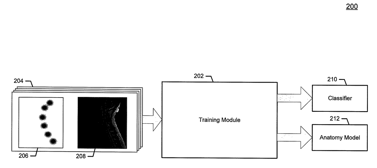
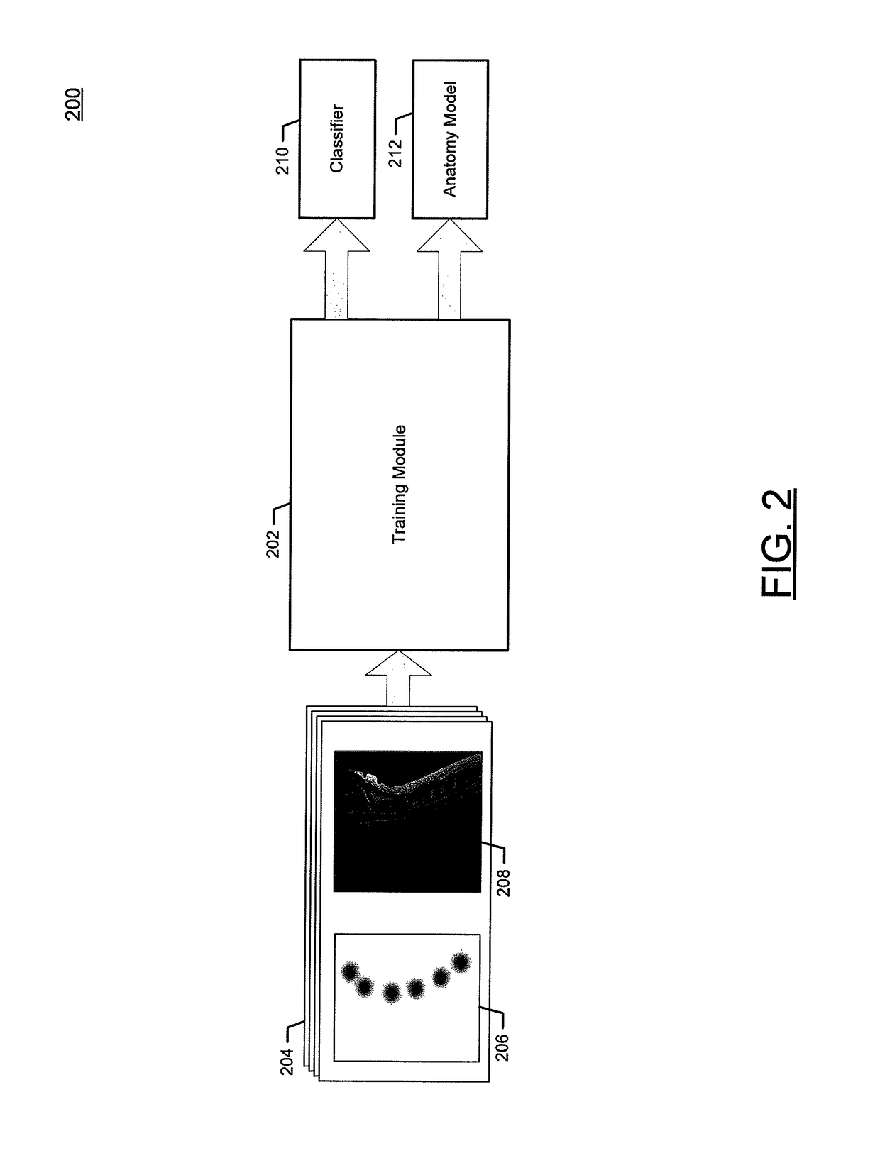
[0032]FIG. 2 is a block diagram of a process flow 200 for generating an image classifier and an anatomical model in accordance with example embodiments of the present invention. The process flow 200 may include a training module 202 that receives a set of training images 204. The set of training images 204 may include a plurality of images, including one or more target images 206 and one or more source images 208. The target images 206 represent a mapping of the location of particular anatomical elements to be detected by a generated image classifier. For example, a target image 206 might include data specifying the location of vertebrae in a spinal image. In the present example, the pixels of the target image 206 only display the locations of vertebrae. Each of the target images 206 may be associated with a particular source image 208, representing an image from which the corresponding target image 206 was derived. For example, a target image 206 and source image 208 p...

example labeling

Data Flow

[0034]Turning now to FIG. 3, an example data flow 300 for labeling anatomical elements within a medical image is depicted. The data flow 300 includes a training phase 302, which includes generating one or more image classifiers and an anatomical model. The image classifier is generated and utilized in a feature detection phase 304, and the anatomical model is generated and utilized in a pictorial phase 306. It should be readily appreciated that, while the training phase 302 is shown as overlapping with portions of the feature detection phase 304 and the pictorial phase 306, the training phase 302 could be decoupled from the use of the image classifier and anatomical model to label medical images. For example, the training phase 302 could be performed as part of a calibration or configuration step at a factory or laboratory, and a set of pre-generated image classifiers and anatomical models could be provided along with sale of a product or device used for labeling of anatomi...

example method

for Generating an Anatomical Model

[0080]Turning now to FIG. 12, an example embodiment for generating an anatomical model is described. As described above, an anatomical model may be generated for use in conjunction with an image classifier to detect the location of one or more anatomical elements in a test image. Generation of the anatomical model may be performed using pictorial image evaluation techniques, where the location of particular anatomical elements are derived from known data and a model is generated describing the relationship between these anatomical elements. This model may be used to describe the size, shape, structure, form, location, and the like of the anatomical elements.

[0081]At action 1202, a set of anatomical data may be received. As described above with respect to FIG. 4, the set of anatomical data may include a set of training images that are presented as a set of target images and a set of source images. In some additional or alternative embodiments, the an...


PUM


Abstract

A method, apparatus and computer program product are hereby provided to detect anatomical elements in a medical image. In this regard, the method, apparatus, and computer program product may receive a test image and generate a classified image by applying an image classifier to the test image. The image classifier may include at least one decision tree for evaluating at least one pixel value of the test image and the classified image may include a plurality of pixel values. Each pixel value may be associated with a probability that an anatomical element is located at the pixel location. The method, apparatus, and computer program product may also evaluate the classified image using an anatomical model to detect at least one anatomical element within the classified image.

Description

TECHNOLOGICAL FIELD[0001]An example embodiment of the present invention relates generally to medical imaging applications, and, more particularly, to a method and apparatus for detecting anatomical elements within a medical image.BACKGROUND[0002]As medical technology has advanced, it is increasingly common for practitioners to interpret data received from one or more medical imaging devices. Different types of imaging devices (e.g., x-ray images, computed tomography scans, magnetic resonance imaging (“MRI”) scans) produce different types of output images, and medical practitioners receive extensive training in performing diagnoses based on these images. However, analysis of these images may involve various time consuming processes, such as manual labeling of elements within an image. For example, a practitioner might spend a significant amount of time manually applying labels to vertebrae in a spinal x-ray or MRI scan in order to report a spinal problem. These manual processes may d...

Claims


Application Information

Patent Timeline
Patent Type & Authority Patents(United States)
IPC IPC(8): G06K9/62A61B5/00G06K9/46G06T7/00
CPCG06K9/6224A61B5/7264G06K9/4614G06K9/6282G06T7/0012G06K9/6267G06T2207/20081G06K2209/05G06V10/446G06V2201/03G06V10/7635G06F18/2323G06F18/24G06F18/24323
Inventor REZAEE, MAHMOUD RAMZETOP, ANDREWBROWN, COLINHAMARNEH, GHASSAN
Owner CHANGE HEALTHCARE HLDG LLC
Why Eureka
Try Eureka