Tissue engineering complex grid shape stent forming method base on core dissolving technology

A technology of tissue engineering and mesh scaffolds, which is applied in the field of forming complex tubular mesh scaffolds in tissue engineering, can solve the problems of inability to form spatial mesh structures, complex tubular mesh scaffold forming methods, and limited types of formable materials, etc., to achieve Rapid forming, large processing capacity and wide range of applicable materials

Inactive Publication Date: 2009-05-27
TSINGHUA UNIV
View PDF7 Cites 2 Cited by
  • Summary
  • Abstract
  • Description
  • Claims
  • Application Information

AI Technical Summary

Problems solved by technology

[0013] The present invention aims at the problems of limited formable materials in the current tubular support technology, the inability to form a tubular support with a spatial network structure, and the inability to form a tubular support with a material gradient and a pore gradient in the tube wall, and develops a core-dissolving technology. Tissue Engineering Complex Tube-Network Scaffold Forming Method

Method used


Image

  • Tissue engineering complex grid shape stent forming method base on core dissolving technology
  • Tissue engineering complex grid shape stent forming method base on core dissolving technology
  • Tissue engineering complex grid shape stent forming method base on core dissolving technology

Examples


Embodiment 1

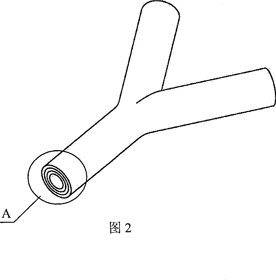
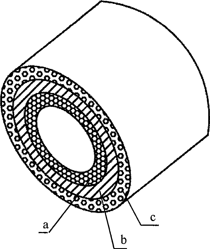
[0035] In a tissue engineering, a tubular network carrier tubular scaffold with a planar bifurcation structure is required. The maximum diameter of the "trunk" is 3mm, and there are two bifurcation structures under the trunk, and the bifurcation diameter is 3mm. The stent tube wall has a three-layer structure. The inner wall needs a microporous structure with a pore size of 70um, the porosity is about 90%, and the material is PLGA. The outer wall needs a microporous structure with a pore size of 120um, the porosity is about 75%, and the material is the same as the inner layer. The middle layer has no microporous structure, and the material is PCL-PET multi-block copolymer.

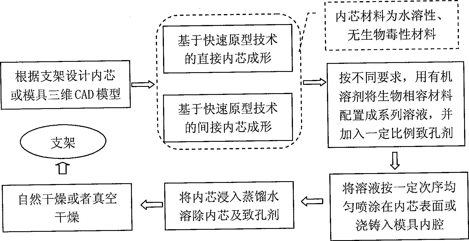
[0036] (1) Design the inner core of the stent. Use SolidWorks 3D CAD software to design the 3D structure of the inner core, and output the CAD model of the stent inner core as STL file format.

[0037] (2) The inner core is made by melt extrusion process (MEM process). The inner core forming material i...

Embodiment 2

[0053] Three-dimensional block scaffolds with inner tube network were prepared. Inside the bracket, there is an inner pipe network that communicates with each other. The inner pipe network of the stent has a three-layer space bifurcation structure and four-level pipe diameters, and the diameters are 1200 μm, 900 μm, 600 μm and 300 μm in sequence. The shape of the inner pipe network is as image 3 shown. The manufacturing process of the bracket is as follows:

[0054] (1) According to the shape of the inner pipe network, use Solidworks software to design the inner core of the inner pipe network. According to the appearance and shape of the stent, design the outer contour and inner core of the stent. And output the resulting models as STL files respectively. The inner core shape of the inner pipe network is as follows Figure 4 shown

[0055] (2) The inner core of the inner pipe network is indirectly formed based on the stereolithography process.

[0056] First, the inne...

Embodiment 3

[0066] A common bile duct stent with a planar bifurcated structure was prepared. The macroscopic structure of the stent is a main tube with an inner diameter of 9mm, and two bifurcations with an inner diameter of 8mm and 6mm are divided into two faces. The tube wall requires no microporous structure, and the stent forming material is PLLGA.

[0067] (1) Design the inner core of the stent. Use SolidWorks 3D CAD software to design the 3D structure of the inner core, and output the CAD model of the stent inner core as STL file format.

[0068] (2) Direct forming of inner core based on melt extrusion process.

[0069] The stent outer contour support was fabricated using the MEM250-II melt extrusion molding system produced by Beijing Yinhua Laser Rapid Prototyping and Die Technology Co., Ltd. The forming material is a mixture of maltose, glucose and sucrose. The main process parameters used are: layer thickness 0.20mm, scanning speed 23mm / s.

[0070] (3) 1 g of PLLGA (Mv: 1000...


PUM

PropertyMeasurementUnit
diameteraaaaaaaaaa
elastic modulusaaaaaaaaaa
porosityaaaaaaaaaa

Abstract

The method of complicated tubular netted tissue engineering rack forming technology based on core dissolving technique belongs to the field of tissue engineering rack manufacturing technology. The technological process includes first designing 3D model of rack and rack core; making rack core with water soluble material without biotoxicity through fast laminated formation; compounding solution of biocompatible material with proper amount of pore creating agent and coating the solution onto the rack core; air drying, soaking in distilled water to dissolve out rack core and pore creating agent, taking out and volatilizing water and coating different forming material with or without pore creating agent successively to form the tubular netted rack with complicated spatial structure, different material gradient and different pore gradient.

Description

technical field [0001] The invention relates to a method for forming a complex tubular network stent for tissue engineering, and belongs to the technical field of forming and manufacturing of tissue engineering stents. Background technique [0002] As one of the three elements of tissue engineering, tissue engineering scaffolds provide the material basis for cell growth and mechanical support, and are the basis for tissue engineering research. Human tissue engineering technology requires the scaffold to have good biocompatibility and degradability; appropriate mechanical properties; macroscopic morphology similar to the target organ and certain microporous structure. The macroscopic shape of the scaffold basically determines the final shape of the artificial tissue or organ. In the tissue engineering of nerve conduit repair, blood vessels, salivary glands, tissue engineering and other organs with hollow tubular as the main functional structure, such as digestive tract, resp...

Claims


Application Information

Patent Timeline
Patent Type & Authority Patents(China)
IPC IPC(8): A61F2/00A61F2/04A61F2/06A61L27/00
Inventor 林峰颜永年张磊卢清萍王小红熊卓吴任东张人佶向虎陶梅
Owner TSINGHUA UNIV
Why Eureka
Try Eureka