Nuclear medical imaging device

An image and x-ray technology, applied in the field of medical imaging, can solve problems such as parallel processing and slow acquisition of gamma images

Inactive Publication Date: 2007-11-07
杰克·E·朱尼
View PDF4 Cites 5 Cited by
  • Summary
  • Abstract
  • Description
  • Claims
  • Application Information

AI Technical Summary

Problems solved by technology

However, x-ray imaging is generally a faster procedure compared to SPECT imaging, and there are no prior devices that allow x-ray and gamma imaging to be performed in parallel
Often, x-ray images are acquired quickly, followed by gamma images relatively slowly, leading to serious problems in data correlation

Method used


Image

  • Nuclear medical imaging device
  • Nuclear medical imaging device
  • Nuclear medical imaging device

Examples


Embodiment Construction

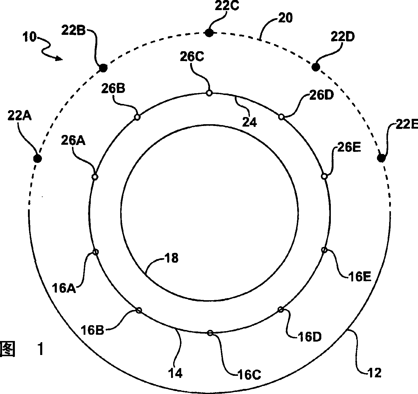
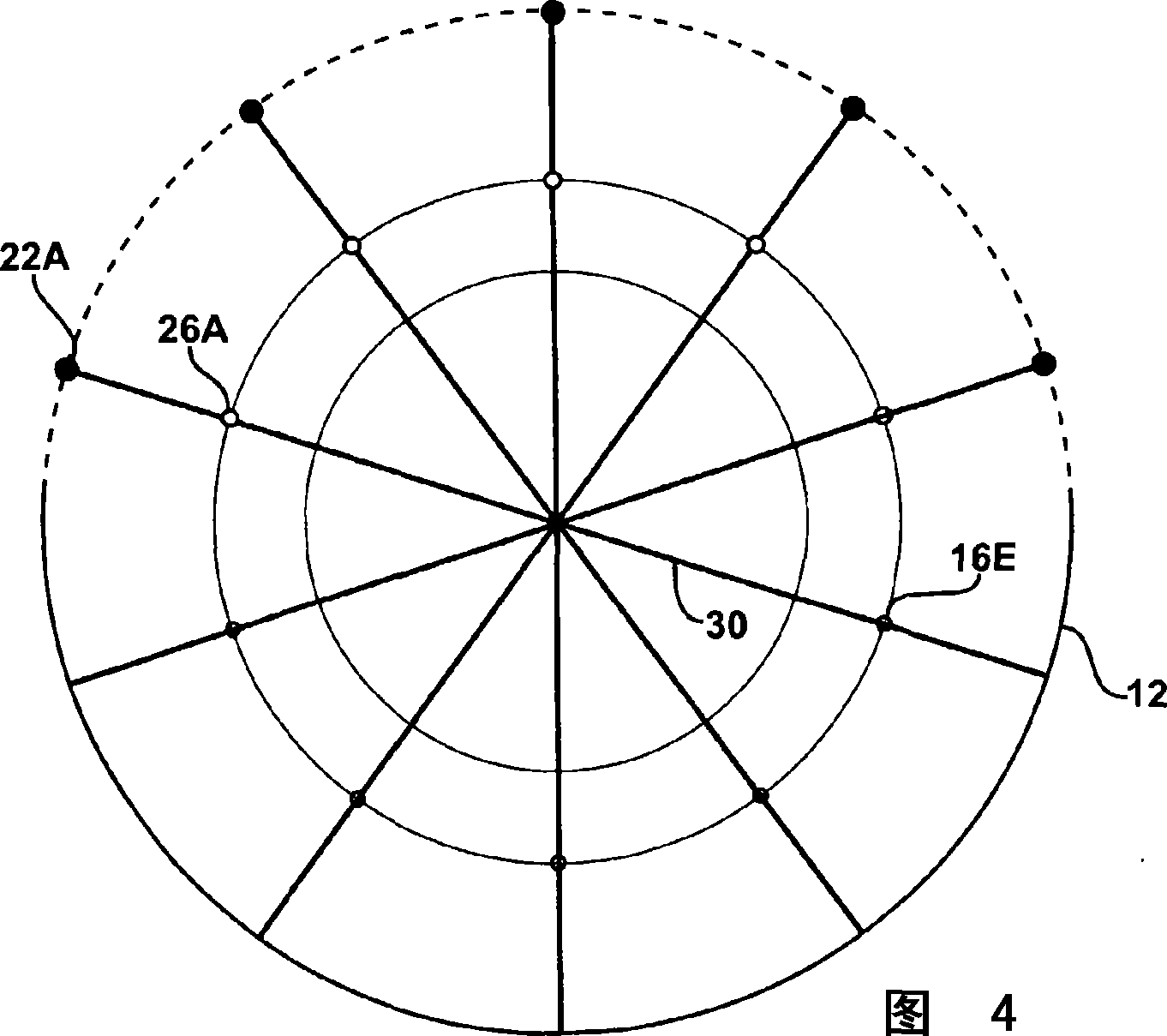
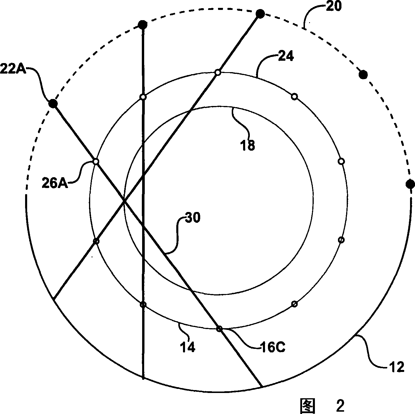
[0038] Embodiments of the present invention provide improved imaging of objects. Imaging can be performed using both electromagnetic wavelength regions, eg using x-ray imaging and gamma imaging (such as SPECT). X-ray attenuation measurements can be used to provide improved SPECT images by providing correction for the attenuation variability of gamma photons within the object. Attenuation coefficients may be assumed to be the same for gamma photons or x-ray photons, or an object model used to convert between attenuation coefficients for attenuation correction.

[0039] In an exemplary embodiment of the invention, attenuation measurements are made by determining the attenuation of a radiation beam passing through the subject. The radiation source is located close to the field of view, and the radiation from the source is detected after passing through the field of view. By changing the direction of the path of radiation through the field of view, a radiation transmission (atte...


PUM

PropertyMeasurementUnit
radiusaaaaaaaaaa
radiusaaaaaaaaaa

Abstract

An apparatus for imaging a field of view, according to an embodiment of the present invention, comprises an x-ray source positioned adjacent to the field of view, a source mask located between the x-ray source and the field of view having at least one source aperture defined therethrough, and an emission detector, such as a gamma camera, adjacent to the field of view. The apparatus may allow substantially concurrent x-ray imaging and gamma imaging of the field of view.

Description

[0001] References to related applications [0002] This application claims priority from U.S. Provisional Patent Application Serial No. 60 / 607,319, filed September 3, 2004, and Serial No. 60 / 652,424, filed February 11, 2005 US Provisional Patent Application, both of which are hereby incorporated by reference in their entirety. technical field [0003] The present invention relates generally to imaging devices and imaging methods, and more particularly to medical imaging. Background technique [0004] Medical imaging devices come in many forms, including nuclear medicine imaging devices such as those described in Applicant's co-pending patent application, which is hereby incorporated by reference in its entirety. As is known to those of ordinary skill in the art, when using nuclear medicine imaging equipment, such as SPECT equipment, it is desirable to determine the amount of radiation attenuation caused by portions of the subject surrounding the imaged region. [0005] SPE...

Claims


Application Information

Patent Timeline
Patent Type & Authority Applications(China)
IPC IPC(8): G01N23/06G01F1/66
Inventor 杰克·E·朱尼
Owner 杰克·E·朱尼
Why Eureka
Try Eureka