Method for removing improved conical bind CT ring shaped false shadow

A ring artifact and cone beam technology, applied in the field of medical CT image processing, can solve problems such as misjudgment, achieve accurate artifact recognition, high resolution, and eliminate ring artifacts

Inactive Publication Date: 2008-05-14
SOUTHERN MEDICAL UNIVERSITY
View PDF1 Cites 41 Cited by
  • Summary
  • Abstract
  • Description
  • Claims
  • Application Information

AI Technical Summary

Problems solved by technology

This method defines the filter in the direction of the vertical axis, and uses 2.5 times the sum of the pixel values ​​of the two points at the two ends of the 7 pixels under the filter to subtract the "difference" of the sum of the pixel values ​​​​of the center 5 points. Compare with the upper and lower limits set by the system, and then judge whether there is an artifact in the center pixel of the filter. Therefore, when the width of the artifact line is greater than or equal to the width of 7

Method used


Image

  • Method for removing improved conical bind CT ring shaped false shadow
  • Method for removing improved conical bind CT ring shaped false shadow
  • Method for removing improved conical bind CT ring shaped false shadow

Examples


Embodiment Construction

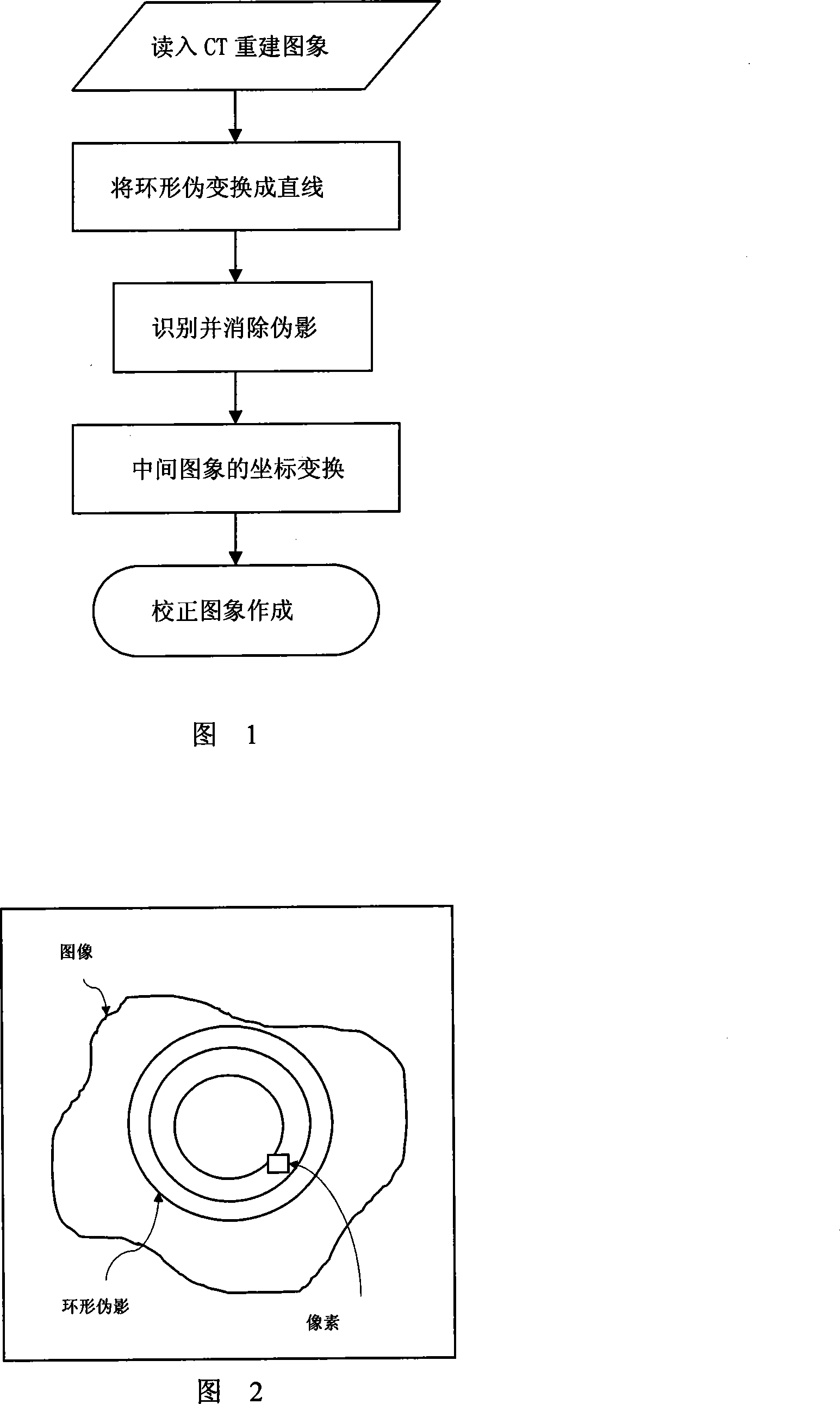
[0033] The method of the present invention will be described in detail below in conjunction with the accompanying drawings and a set of cone-beam CT images of mice reconstructed with the FDK algorithm. The specific steps are as follows:

[0034] Step 1: Read in the cone-beam CT image of the mouse reconstructed by the FDK algorithm, the size of which is 512×256, and convert the grayscale image into a floating-point image through linear transformation, which is suitable for coordinate transformation. The specific calculation formula is: I-I min / (I max -I min ), where I is the image pixel value, I max and Imin They are the maximum and minimum grayscale values ​​of the image to be processed, which are 233 and 0 in this example.

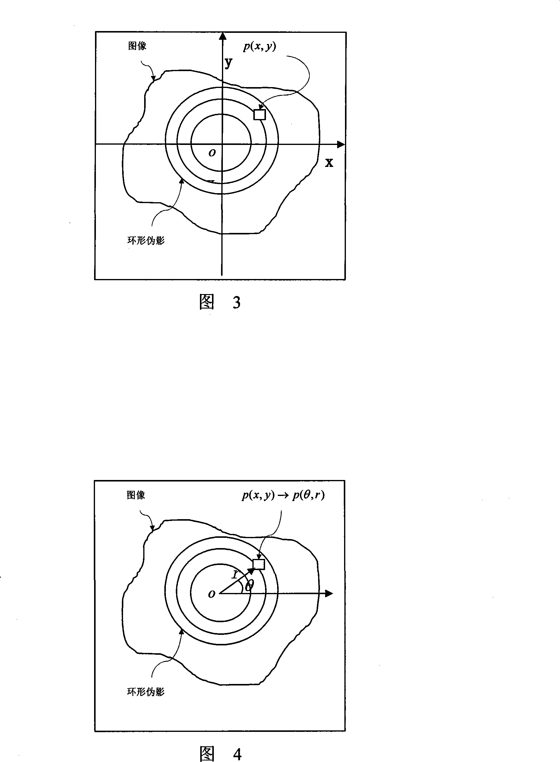
[0035] Step 2: Perform coordinate transformation (see Figures 2-4). The origin o of the rectangular coordinate system where the CT image is located is the pole o of the polar coordinate system, and the horizontal axis x of the rectangular coordinate...


PUM


Abstract

The invention discloses an improved eliminating method of tapered beam CT reconstruction image annular false shade, comprising the steps as follows: the invention changes the annular false shade of reading CT reconstruction image into a straight line through a method of coordinate change, then takes a one-dimension filter parallel with the false shade as a mask to search a middle image regularly line by line and acquire sample space, at the same time calculates out the variance about the pixel value of each sample space, acknowledges the false shade according to whether the variance is less than the preset valve value, substitutes the pixel value of each sample space with the average pixel value between the point and two upper and lower false shades in the ordinate axis direction in order to eliminate the annular false shade, and finally changes the middle image free from the false shade to be under the original frame of axes to gain a corrected image.

Description

technical field [0001] The invention relates to a processing method of medical CT images, in particular to a method for eliminating ring artifacts in cone-beam CT reconstruction images. Background technique [0002] Computed tomography, or CT (Computed Tomography), has been widely used in the field of clinical medicine in the 20th century. Since the birth of CT, its scanning method has developed from a two-dimensional single-detector parallel beam to a multi-detector fan-beam rotary scanning, and a three-dimensional CT has developed from the earliest single-row spiral CT to multi-row detector scanning. -Multislice spiral / helical CT (MSCT) with 32 layers of data. In recent years, cone-beam CT (CBCT) using area array detectors has gradually entered the practical stage. It uses cone-beam scanning to obtain two-dimensional projection data at various viewing angles, and directly obtains three-dimensional volumes after reconstruction. Compared with other geometric structures, th...

Claims


Application Information

Patent Timeline
IPC IPC(8): G06T5/00G06T11/00A61B6/03
Inventor 甄鑫周凌宏王卓宇张书旭卢文婷陈超敏
Owner SOUTHERN MEDICAL UNIVERSITY
Why Eureka
Try Eureka