Method and apparatus for three-dimensional visualization of sequence image

A sequential image and three-dimensional technology, applied in the field of medical image processing, can solve the problems of slow imaging speed and inconvenient operation of three-dimensional visualization operation, and achieve the effect of fast and convenient imaging, convenient establishment, and less manual participation

Inactive Publication Date: 2008-09-03
SOUTH CHINA NORMAL UNIVERSITY +1
View PDF0 Cites 20 Cited by
  • Summary
  • Abstract
  • Description
  • Claims
  • Application Information

AI Technical Summary

Problems solved by technology

[0004] In the existing three-dimensional visualization operation of medical image processing, two-dimensional image segmentation is usually performed on a group of sequence images of organs or tissues. In one of the image segmentation methods, when segmenting sequence images, It is to divide the sequential images into groups of six, and each group shares an artificial initialization contour. After the image segmentation, the 3D reconstruction operation is performed to realize the 3D visualization process. In the existing 3D visualization processing method, it is necessary to Initializing the outline of each group of pictures obviously adds more manual intervention operations. In addition, since two-dimensional image segmentation is only performed on a two-dimensional plane, and organs or tissues are three-dimensional objects, when represented by sequence images, in which On an image of the organ or tissue, the area or range displayed by the organ or tissue will be relatively small, and the two-dimensional operation needs to manually initialize the outline for each image, which will easily lead to slow and inconvenient imaging in the three-dimensional visualization operation

Method used


Image

  • Method and apparatus for three-dimensional visualization of sequence image
  • Method and apparatus for three-dimensional visualization of sequence image
  • Method and apparatus for three-dimensional visualization of sequence image

Examples


Embodiment 1

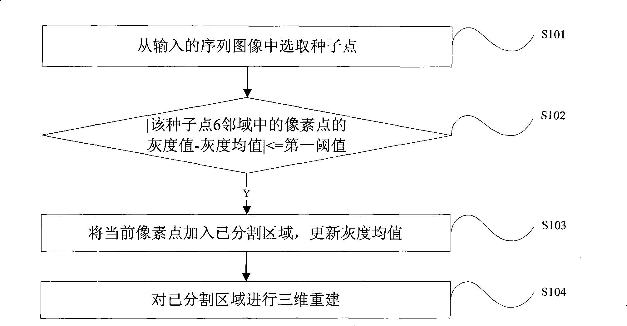
[0021] like figure 1 As shown, it is a schematic flowchart of Embodiment 1 of the method for 3D visualization of sequence images according to the present invention, which includes the steps:

[0022] Step S101: arbitrarily select a slice image containing the required organ or tissue data from the input sequence image, and select a seed point from the slice image;

[0023] Step S102: Obtain the grayscale value of each pixel in the six neighborhoods of the seed point, and judge the absolute value of the difference between the grayscale value of each pixel and the average grayscale value of the segmented area, and determine the absolute value. Whether it is less than or equal to the first threshold, when the determination result is no, the current pixel is not processed, that is, the pixel is not added to the segmented area, wherein the first threshold can be manually set according to experience;

[0024] Step S103: when the determination result of the step S102 is yes, add the ...

Embodiment 2

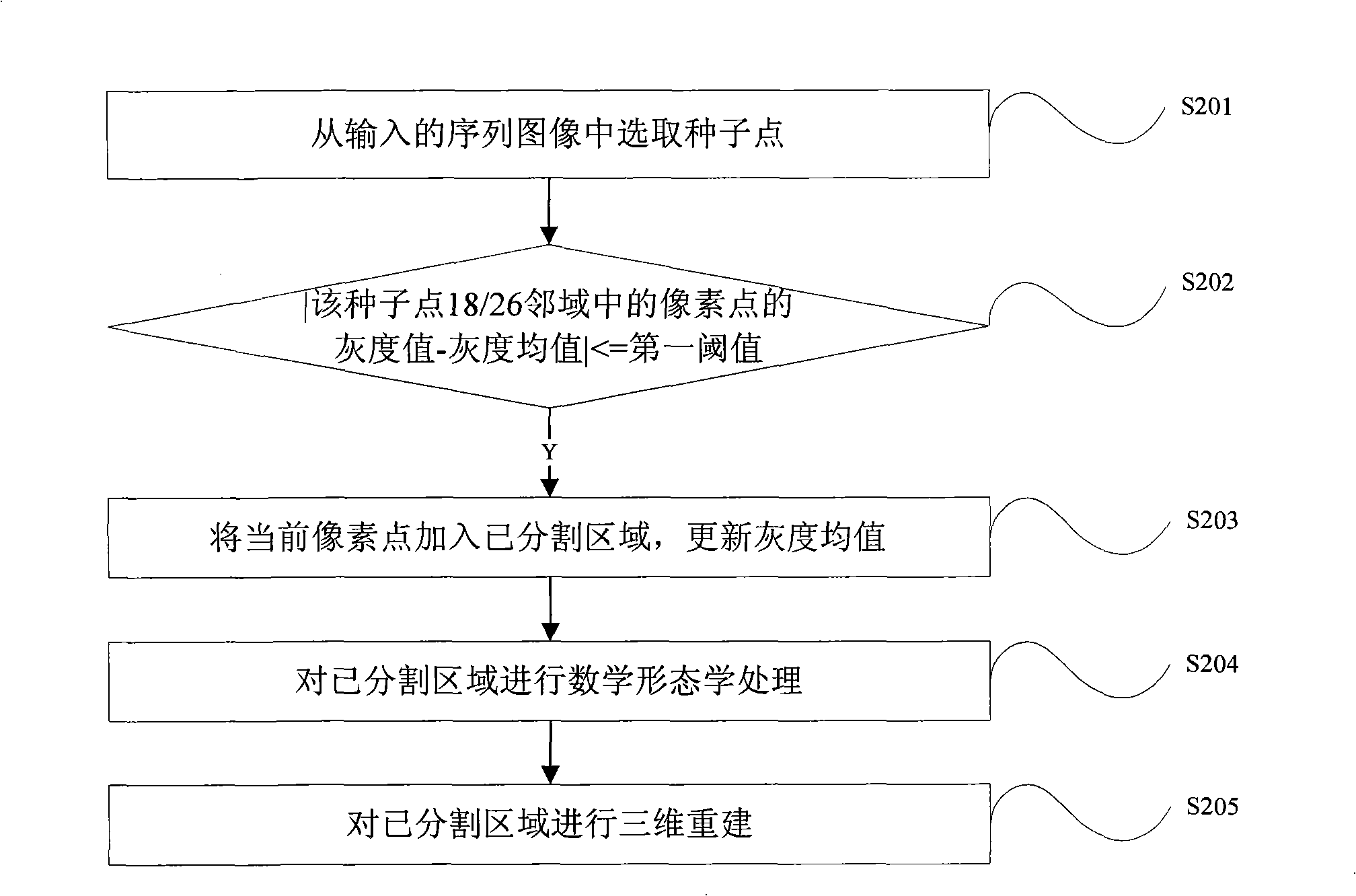
[0037] like figure 2 shown is a schematic flowchart of Embodiment 2 of the method for 3D visualization of sequence images according to the present invention. In this embodiment, the main difference from Embodiment 1 lies in that, before performing 3D reconstruction on the pixels of the segmented area, it also includes: The steps of performing mathematical morphological processing on the pixels of the segmented area are presented. In this embodiment, it specifically includes steps:

[0038] Step S201: arbitrarily select an image containing the required organ or tissue data from the input sequence images, and select seed points from the image;

[0039] Step S202: Obtain the grayscale value of each pixel in the eighteenth neighborhood or twenty-six neighborhood of the seed point, and determine the difference between the grayscale value of each pixel and the average grayscale value of the segmented area. Absolute value, determine whether the absolute value is less than or equal t...

Embodiment 3

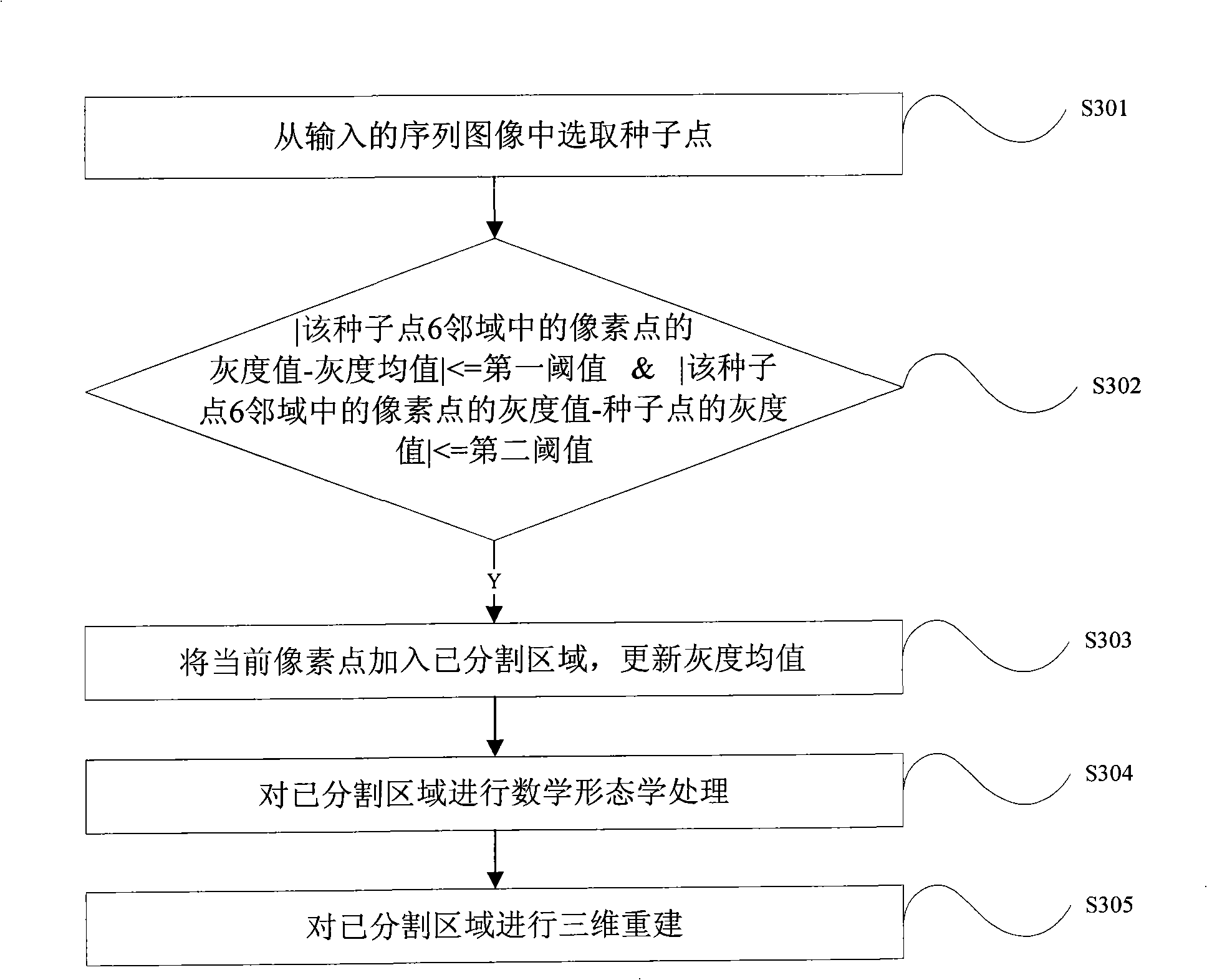
[0047] like image 3 shown is a schematic flowchart of Embodiment 3 of the method for 3D visualization of sequence images according to the present invention. In this embodiment, the difference from Embodiment 2 is that when judging whether a pixel can be added to a segmented area, increase A new judgment condition is set. It specifically includes steps:

[0048] Step S301: arbitrarily select an image containing the required organ or tissue data from the input sequence images, and select seed points from the image;

[0049] Step S302: Obtain the gray value of each pixel in the six neighborhoods of the seed point, and judge the absolute value of the difference between the gray value of each pixel and the average gray value of the divided area, and determine the absolute value. Whether it is less than or equal to the first threshold, and at the same time judge whether the absolute value of the difference between the gray value of the pixel point and the gray value of the seed p...


PUM


Abstract

The invention relates to a method and a device for sequence image three-dimensional visualization, which chooses a seed point from input sequence image, adds the pixel to a divided region when the absolute value of the difference value between the gray value of the pixel in the three-dimensional neighborhood of the seed point and the mean gray of the divided region is less than or equal to a first threshold value, then carries out three-dimensional reconstruction with the pixels in the divided region. According to the method and device for sequence image three-dimensional visualization of the invention, which chooses a seed point from sequence image, determines whether the absolute value of the difference value between the gray value of the pixel in the three-dimensional neighborhood of the seed point and the mean gray of the divided region is less than or equal to the first threshold value, and adds the pixel to the divided region if it is true. Since the invention carries out the comparison based on the mean gray of the divided region, and carries out the judgment in the three-dimensional region of the seed pixel point, thus it is easier to construct the three-dimensional model for image, it needs few persons to take part in, and it is rapid and convenient for imaging.

Description

technical field [0001] The invention relates to the field of medical image processing, in particular to a method and device for three-dimensional visualization of sequence images in the field of medical image processing. Background technique [0002] Image segmentation refers to decomposing an image into a set of several non-overlapping regions. The result of image segmentation determines the quality of the final output in various applications in advanced vision. In the field of medical images, the purpose of image segmentation is is to efficiently identify clinically morphological information that is critical to the patient for further analysis. The three-dimensional reconstruction refers to the restoration of the three-dimensional result of the object from the acquired sampling data, that is, the prototype of the object. [0003] In the medical field, it is usually necessary to obtain a set of tomographic images inside the human body through various medical equipment, and...

Claims


Application Information

Patent Timeline
Patent Type & Authority Applications(China)
IPC IPC(8): G06T5/00G06T11/00G06T17/00A61B19/00A61B90/00
Inventor 鲍苏苏方驰华廖其光黄燕鹏庞雄文
Owner SOUTH CHINA NORMAL UNIVERSITY
Why Eureka
Try Eureka