Method for partitioning two-dimensional sequence medical image based on prior knowledge earth-measuring geometry flow

A two-dimensional sequence, prior knowledge technology, used in image analysis, image data processing, instrumentation, etc.

Inactive Publication Date: 2008-11-12
HARBIN INST OF TECH
View PDF0 Cites 13 Cited by
  • Summary
  • Abstract
  • Description
  • Claims
  • Application Information

AI Technical Summary

Problems solved by technology

But since this term only depends on the gradient information of the image, this external force may still not be enough to completely solve the problem of gap leakage at weak edges of the image

Method used


Image

  • Method for partitioning two-dimensional sequence medical image based on prior knowledge earth-measuring geometry flow
  • Method for partitioning two-dimensional sequence medical image based on prior knowledge earth-measuring geometry flow
  • Method for partitioning two-dimensional sequence medical image based on prior knowledge earth-measuring geometry flow

Examples


Embodiment Construction

[0036] Below in conjunction with accompanying drawing and specific embodiment the present invention will be described in further detail:

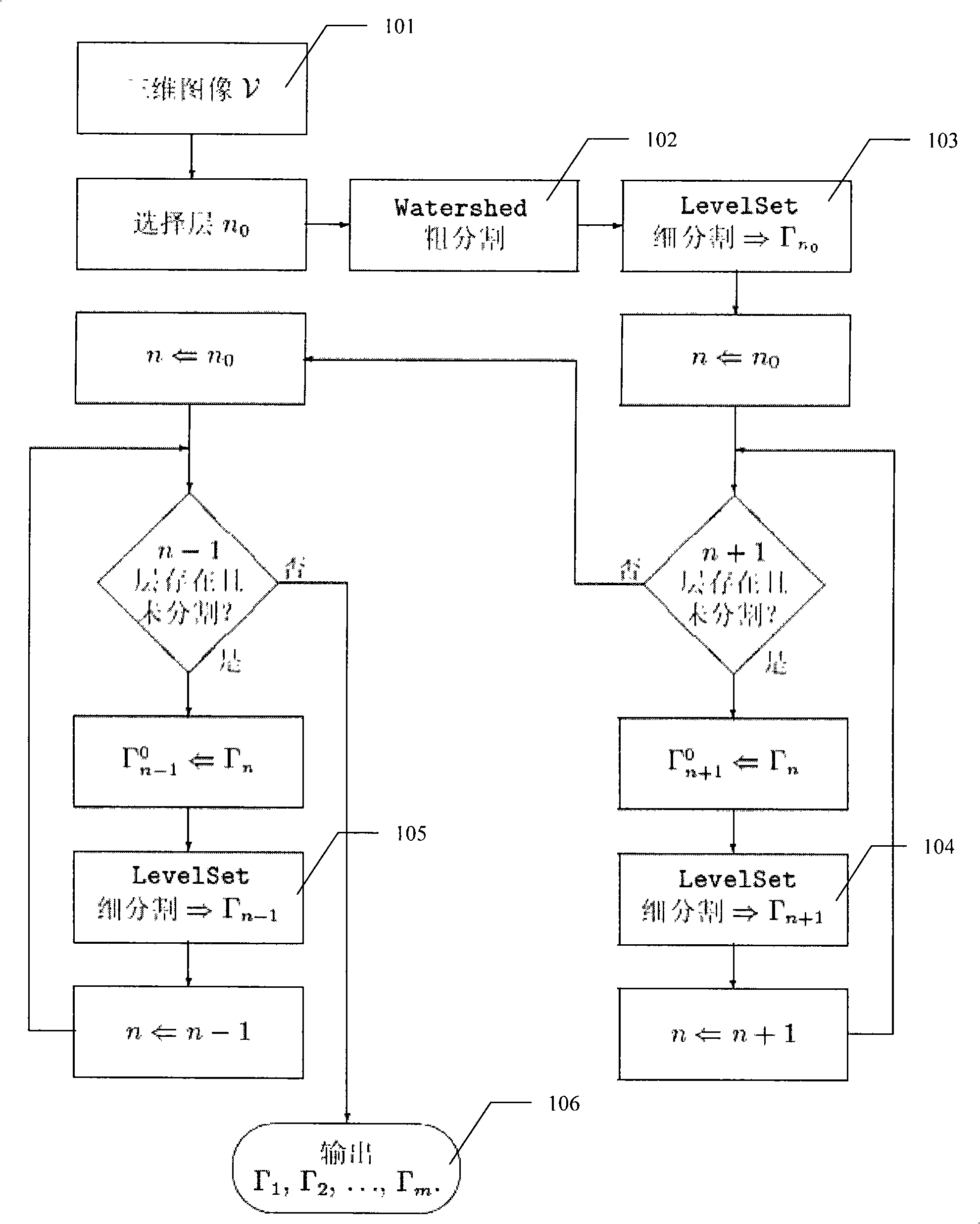
[0037] The method involved in the present invention is realized by computer numerical calculation. The implementation is mainly divided into two parts. One is to select an appropriate initial layer and obtain the segmentation result, and the other is to use the previous segmentation result as a priori reference information to segment the next adjacent layer. For specific procedures, see figure 1 . figure 1 Middle: first select layer n of the two-dimensional sequence 101 of the original three-dimensional brain MR image 0Carry out rough watershed segmentation 102, in which the traditional geodesic geometric flow fine segmentation 103 is firstly performed, and the rough segmentation result of the initial layer is used as the initial boundary, and then the adjacent layer images on the upper and lower sides from the initial layer are refined on...


PUM


Abstract

The invention provides a two-dimensional sequence medical image segmentation method based on prior knowledge geodesic geometric flow. Firstly, a layer of two-dimensional image is initially segmented by applying Watershed Algorithm and the rough segmentation result of the initial layer is taken as an initial boundary; subsequently, a resegmentation is performed to adjacent layers of images at the upper lower sides respectively from the initial layer layer by layer in a level set method; wherein, the resegmentation result of each layer is taken as the initial boundary of the next layer and the gradient prior knowledge information is provided to the next layer in the form of adjacent layer gradient reference term for segmentation layer by layer till all of the layers are segmented; finally, the segmentation results of all the layers are combined. By introducing the prior knowledge of the adjacent layer in an edge detection function to improve the stop condition of curve evolution and introducing geodesic geometric flow by taking interlayer gradient similarity as the prior knowledge, the method improves the phenomena of edge leakage when a geometric active contour model is opposite to the discontinuous edge or weak edge in the layers and enhances the precision and stability of three-dimensional medical image segmentation.

Description

(1) Technical field [0001] The invention relates to the field of medical image segmentation, in particular to an efficient three-dimensional medical image segmentation method. (2) Background technology [0002] In medical image processing and analysis applications, image segmentation technology plays a key role. The task of medical image segmentation is to extract the region of interest (Region of Interest) containing important diagnostic information from medical images, so as to provide a reliable basis for clinical diagnosis and pathology research. Due to the complexity and difference of the imaging principle of medical images and the structure of human tissue itself, medical images inevitably have the characteristics of blur and inhomogeneity compared with ordinary images; at the same time, the rapid development of medical imaging technology makes various complex images Massive medical image data has become possible, all of which put forward higher requirements for segme...

Claims


Application Information

Patent Timeline
Patent Type & Authority Applications(China)
IPC IPC(8): G06T7/60
Inventor 沈毅王强郝家胜
Owner HARBIN INST OF TECH
Why Eureka
Try Eureka