Multimodal imaging system for tissue imaging

A tissue and imaging technology, applied in the fields of application, diagnosis with light, medical science, etc., can solve problems such as OCT scanning

Inactive Publication Date: 2009-05-20
CARESTREAM HEALTH INC
View PDF1 Cites 31 Cited by
  • Summary
  • Abstract
  • Description
  • Claims
  • Application Information

AI Technical Summary

Problems solved by technology

However, while fluorescence is sensitive to very early tissue lesions, it is also sensitive to bacteria, food, and spots
So fluorescence images often lead to unnecessary OCT scans
[0009] Although many methods and devices have been proposed to combine regional imaging with OCT scanning, there is still much room for improvement

Method used


Image

  • Multimodal imaging system for tissue imaging
  • Multimodal imaging system for tissue imaging
  • Multimodal imaging system for tissue imaging

Examples


Embodiment Construction

[0034] The following description refers in particular to the constituent elements or the more directly associated elements of the components of the device according to the invention. For elements not specifically shown or described, it is to be understood that they may take various forms well known to those skilled in the art.

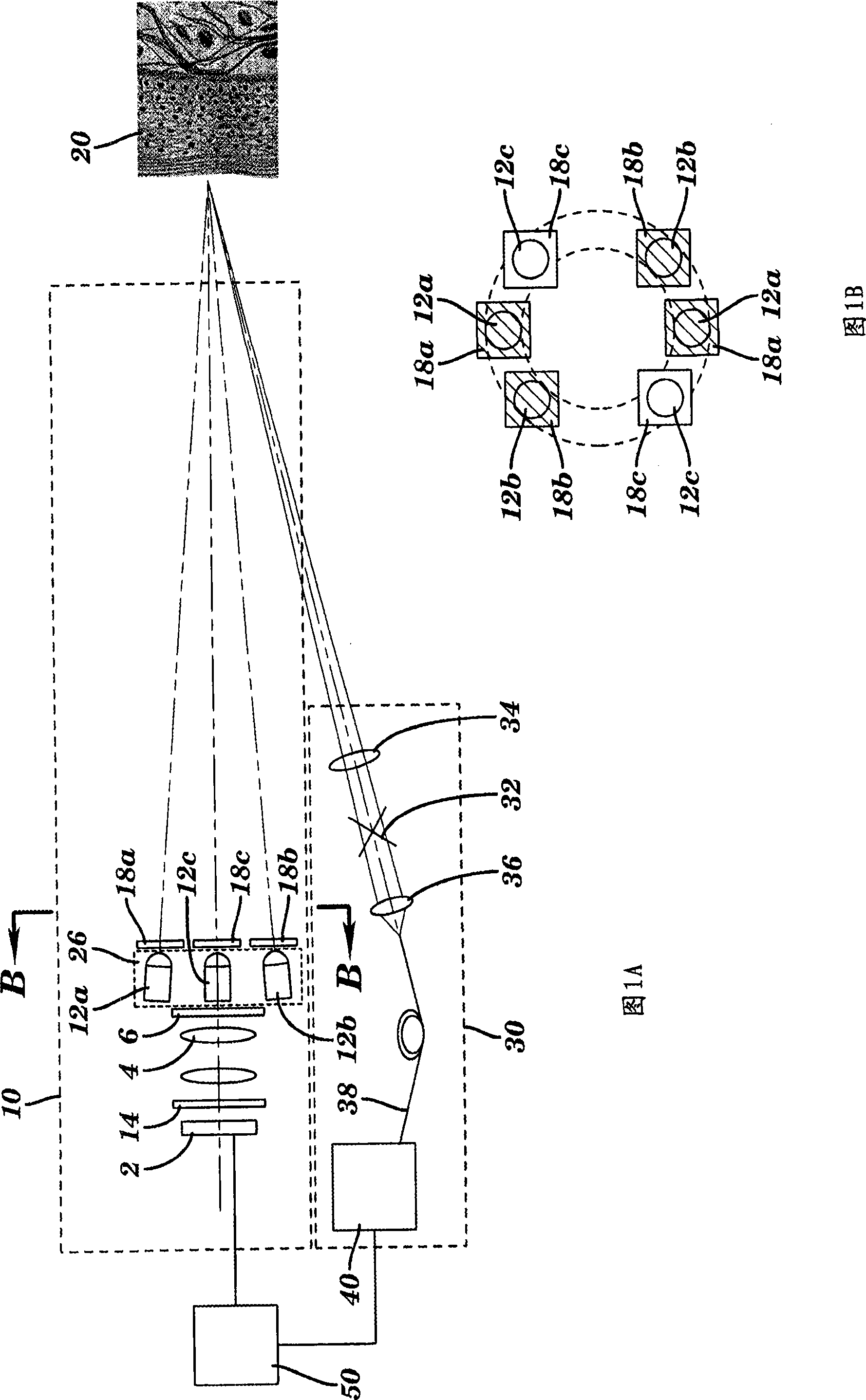
[0035] The present invention combines regional imaging functions (i.e., polarized reflection imaging and fluorescence imaging) with OCT imaging functions. The regional imaging functions are used to identify regions of interest on the tissue surface. For OCT scan data, this particular portion corresponds to a portion of the region of interest.

[0036] Figures 1a and 1b illustrate an imaging device comprising an area imaging system 10 and an OCT imaging system 30 according to one embodiment of the present invention. As part of imaging system 10, light emitting devices 12a, 12b, and 12c provide uniform illumination of the tissue surface for area imaging...


PUM


Abstract

An apparatus for obtaining area images and optical coherence tomography (OCT) image of a tissue comprises an image sensor, a first illumination means providing broadband polarized polychromatic light, a second illumination means providing narrow-band ultraviolet light, a third illumination means providing Near-Infrared light, an optical coherence tomography (OCT) imaging apparatus, one or more polarization elements, and an optical filter. An image processor identifies a region of interest according to area image, which can be a polarized reflectance image, a fluorescence light image, or both for the OCT imaging apparatus to obtain an OCT image over the region of interest.

Description

[0001] Cited Related Patent Applications [0002] U.S. Application Serial No. 11 / 262869, October 31, 2005, titled: Caries Detection Methods, Wong et al.; US Application Serial No. 11 / 408360, April 21, 2006, titled: Optical Detection Methods for Dental Caries, Wong et al.; US Application Serial No. 11 / 530987, September 12, 2006, titled: Caries Detection Device, Liang et al.; US Application Serial No. 11 / 530913, September 12, 2006, titled: Low-Coherence Dental OCT Imaging , Liang et al; U.S. Application Serial No. 11 / 561971, Nov. 21, 2006, titled: Dental OCT Imaging Device, Liang et al; The disclosure of the above co-pending U.S. patent application is incorporated herein. technical field [0003] The present invention generally relates to methods and apparatus for tissue imaging using polarized visible light, ultraviolet light, polarized near-infrared light, and optical coherence tomography (OCT). Background technique [0004] Common methods of histological examination and d...

Claims


Application Information

Patent Timeline
Patent Type & Authority Applications(China)
IPC IPC(8): A61B5/00
CPCA61B5/0059A61B5/0066
Inventor R·梁
Owner CARESTREAM HEALTH INC
Why Eureka
Try Eureka