
Anatomical modeling from a 3-d image and surface mapping

An image and 3-D technology, applied in the field of medical imaging systems, can solve problems such as good resolution

Active Publication Date: 2009-06-10
BIOSENSE WEBSTER INC
View PDF17 Cites 22 Cited by
  • Summary
  • Abstract
  • Description
  • Claims
  • Application Information

AI Technical Summary

Problems solved by technology

Ultrasound imaging is a relatively convenient real-time imaging modality, however the resolution of real-time ultrasound imaging is often not as good as that obtained from other imaging modalities such as computed tomography (CT) and magnetic resonance imaging (MRI)

Method used


Image

  • Anatomical modeling from a 3-d image and surface mapping
  • Anatomical modeling from a 3-d image and surface mapping
  • Anatomical modeling from a 3-d image and surface mapping

Examples


Embodiment Construction

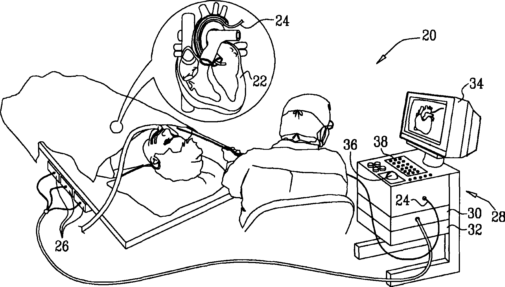
[0067] figure 1 is a schematic illustration of a system 20 for imaging and mapping a target structure (eg, a patient's heart 22 ) in accordance with an embodiment of the present invention. (Hereafter, the term "target structure" may refer to all or part of a ventricle, or to other body cavities, or to a specific wall, surface, vessel, or other anatomical feature. Although the embodiments described herein specifically refer to surrounding structures, but the principles of the invention apply similarly mutatis mutandis to the imaging of bones, muscles and other organs and anatomical structures.)

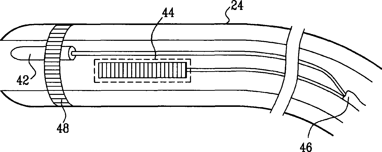
[0068] The system includes a catheter 24, which is inserted into the ventricle by a physician. Typically, catheter 24 is a location-sensing ultrasound probe configured to perform functions including anatomical mapping and ultrasound imaging. The mapping and ultrasound imaging capabilities of catheter 24 are further described in the aforementioned US Patent Publications 2006 / 0253024,...


PUM


Abstract

The invention relates to an anatomical modeling from a 3-D image and a surface mapping. A method of medical imaging includes creating an anatomical map of an inner wall of a cavity in a body of a subject by inserting a probe into the body and collecting data using the probe. A three dimensional (3-D) contour is delineated in a 3-D image of the cavity based on the map.

Description

technical field [0001] The present invention relates generally to medical imaging systems, and in particular to methods and systems for constructing three-dimensional views of internal organs. Background technique [0002] Three-dimensional (3-D) images of internal organs are useful in many catheter-based diagnostic and therapeutic applications, and real-time imaging is widely used during surgical procedures. Ultrasound imaging is a relatively convenient real-time imaging modality, however the resolution of real-time ultrasound imaging is generally not as good as that obtained from other imaging modalities such as computed tomography (CT) and magnetic resonance imaging (MRI). [0003] US Patent 6,106,466 to Sheehan et al., the disclosure of which is incorporated herein by reference, describes a method for generating a 3-D model of the heart from 2-D ultrasound images. Anatomical locations are manually identified in the 2-D images. A 3-D mesh was used as the prototype heart...

Claims


Application Information

Patent Timeline
Patent Type & Authority Applications(China)
IPC IPC(8): A61B8/13A61B5/055A61B6/03A61B19/00
CPCA61B6/541A61B5/1076A61B2019/5289A61B5/7285A61B5/06A61B8/483A61B6/504G06T2207/20156A61B8/0891G06T2207/10072A61B8/4472A61B8/12G06T19/00G06T7/0083A61B8/4488A61B6/5247A61B6/503G06T2207/20136A61B8/5238G06T2207/30048A61B5/042A61B8/0883A61B2090/364G06T7/12G06T7/181A61B5/061A61B5/283
Inventor A·C·阿尔特曼A·戈瓦里
Owner BIOSENSE WEBSTER INC
Learn More