Supercharge Your Innovation With Domain-Expert AI Agents!

Visualizing a vascular structure

A vascular and related technology, applied in cardiac catheterization, image enhancement, image analysis, etc., can solve problems such as insufficient blood supply and obstruction of blood passage through arteries

Active Publication Date: 2009-09-23
KONINKLIJKE PHILIPS NV
View PDF0 Cites 4 Cited by
  • Summary
  • Abstract
  • Description
  • Claims
  • Application Information

AI Technical Summary

Problems solved by technology

These clots block the passage of blood through the arteries, which can lead to insufficient blood supply to the arterial vascular tree of the lungs

Method used


Image

  • Visualizing a vascular structure
  • Visualizing a vascular structure
  • Visualizing a vascular structure

Examples


Embodiment Construction

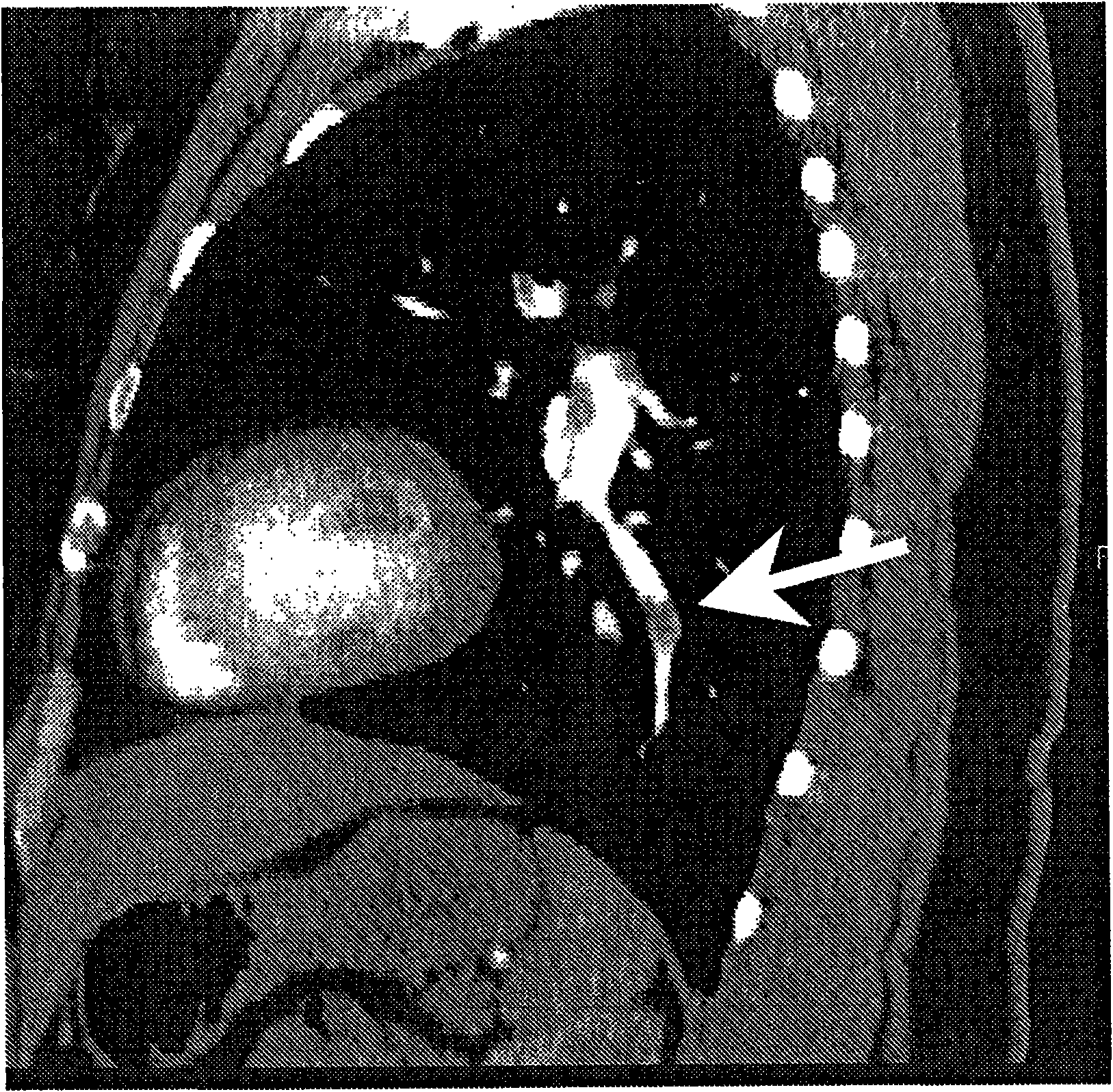
[0042] Pulmonary embolism (PE) is a potentially fatal lung disease involving clots in the pulmonary arteries. These clots block the passage of blood through the arteries, which can lead to insufficient blood supply to the arterial vascular tree of the lung. Clots can be visualized in high-resolution CT volume images due to the absence of contrast agent in the clot resulting in lower Hounsfield values. For example, figure 1 CT slices are shown. The white arrow in the figure points to PE. Visual inspection of pulmonary vessels in initial CT slice images for detection of clots in arteries is often tedious and time consuming. Perfusion defects caused by clots are easily overlooked. Furthermore, it is difficult to quantify the impact of perfusion defects on the entire pulmonary vascular tree by visual inspection of CT images.

[0043] Embolisms also occur in other arteries, including intracranial and coronary arteries. It can also be observed in medical images obtained from o...


PUM


Abstract

A system (500) for visualizing a vascular structure represented by a three- dimensional angiography dataset is disclosed. Respective voxel values are associated with respective voxels. The dataset represents a vascular structure. The system comprises means (502) for establishing respective filling values; means (504) for identifying respective minimum filling values; means (506) for computing respective deficiency values; and an output (514) for providing a visualization in dependence on the deficiency values. A respective filling value is indicative of an amount of blood flow at the respective position in the vascular structure. A respective minimum filling value is a minimum of the filling values associated with the positions upstream of the respective position. A respective deficiency value is indicative of a difference between the filling value associated with the respective position and the minimum filling value associated with the respective position.

Description

technical field [0001] The present invention relates to the visualization of vascular structures, in particular to vascular structures represented by a three-dimensional angiographic data set in which corresponding voxel values ​​are associated with corresponding voxels. Background technique [0002] Pulmonary embolism (PE) is a potentially fatal lung disease involving clots in the pulmonary arteries. These clots block the passage of blood through the arteries, which can lead to undersupply of the arterial vascular tree of the lung. Clots can be visualized in high-resolution CT volume images because of low Hounsfield values ​​due to lack of contrast agent in the clot. [0003] In the article "A novel method for pulmonaryemboli visualization from high-resolution CT images" (Proc. SPIE Medical Imaging 2004), Pichon, Novak, Kiraly, and Naidich describe a method for highlighting potential PEs on a 3D representation of the pulmonary artery tree. First, the pulmonary vessels wer...

Claims


Application Information

Patent Timeline
Patent Type & Authority Applications(China)
IPC IPC(8): A61B6/00G06T7/00G06T11/00
CPCG06T2207/10081G06T7/0012A61B6/032G06T2207/30104A61B6/503A61B6/12A61B6/504A61B6/481
Inventor R·维姆科R·奥普弗T·比洛
Owner KONINKLIJKE PHILIPS NV
Features
  • R&D
  • Intellectual Property
  • Life Sciences
  • Materials
  • Tech Scout
  • Unparalleled Data Quality
  • Higher Quality Content
  • 60% Fewer Hallucinations
Learn More