Device for displaying breast ultrasound information

A breast and equipment technology, applied in the field of breast ultrasound information processing and/or display, can solve the problems of high cost and low work flow efficiency, etc.

Active Publication Date: 2009-10-28
U SYST
View PDF9 Cites 5 Cited by
  • Summary
  • Abstract
  • Description
  • Claims
  • Application Information

AI Technical Summary

Problems solved by technology

Thus, for example, while careful review of raw ultrasound scans by a well-trained radiologist slice-by-slice will improve recall and precision, considering hundreds of individual raw ultrasound slides for each patient, the The overall workflow efficiency of the method will be low and thus the cost will be high

Method used


Image

  • Device for displaying breast ultrasound information
  • Device for displaying breast ultrasound information
  • Device for displaying breast ultrasound information

Examples


Embodiment Construction

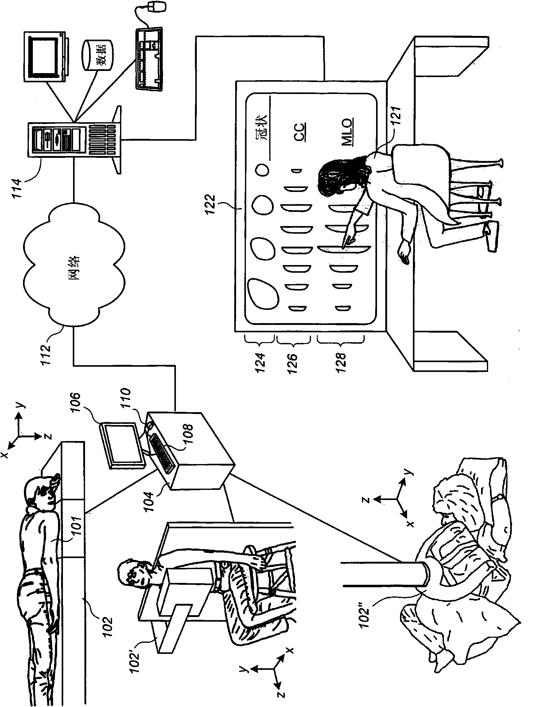
[0047] figure 1 Shown is a conceptual diagram of a breast cancer screening and / or diagnosing system according to a preferred embodiment. The breasts of the patient 101 are ultrasonically scanned with an automated scanning device while the patient 101 is in a prone position (device 102), an upright position (device 102'), a supine position (device 102"), or other positions (not shown). By reducing the required depth of ultrasound penetration into the chest wall, scans of the chest-compressed breast can be performed at higher frequencies such as 10-20 MHz, which can produce images of very high resolution, sufficient to allow detection of volumes of 1 mm near the chest wall However, it should be understood that the scope of the preferred embodiments is not limited to the situation of compression to the chest, where breast ultrasound information processing and display according to the preferred embodiments are generally essential to the process from which the spectral properties o...


PUM


Abstract

Method for treating and displaying breast ultrasound information is disclosed. Displaying breast ultrasound information on an interactive user interface is described, the user interface being useful in adjunctive ultrasound mammography environments and / or ultrasound-only mammography environments. Bilateral comparison is facilitated by a pairwise display of thick-slice images corresponding to analogous slab-like subvolumes in the left and right breasts. Coronal thick-slice imaging and convenient navigation on and among coronal thick-slice images is described. In one preferred embodiment, a nipple marker is displayed the coronal thick-slice image representing a projection of a nipple location thereupon. A convenient breast icon is also displayed including a cursor position indicator variably disposed thereon in a manner that reflects a relative position between the cursor and the nipple marker. Preferably, the breast icon is configured to at least roughly resemble a clock face, the center of the clock face representing the nipple marker location. Bookmark-centric and CAD-marker-centric navigation within and among thick-slice images is also described.

Description

[0001] This application is a patent with the international application number PCT / US2005 / 019604 and the PCT application date of June 1, 2005, after entering the Chinese national phase, the application number is 200580018087.4, and the invention name is "processing and displaying breast ultrasound information" Divisional application of the application. [0002] Cross References to Related Applications [0003] This application claims the benefit of U.S. 10 / 997,293, filed November 23, 2004, provisional application 60 / 577,326, filed June 4, 2004, and provisional application 60 / 577,388, filed June 4, 2004, which Incorporated herein by reference. technical field [0004] This patent specification relates to medical ultrasound imaging. More specifically, this patent specification relates to the processing and / or display of breast ultrasound information for breast cancer screening and / or diagnosis. Background technique [0005] Volumetric ultrasound scanning of the breast can be...

Claims


Application Information

Patent Timeline
Patent Type & Authority Applications(China)
IPC IPC(8): A61B8/08A61B8/13A61B6/02
Inventor 张伟王士平陈驾宇俞增平汤米·厄尔·库普利斯麦克尔·E·里德
Owner U SYST
Why Eureka
Try Eureka