Product for treating myocardial infarction based on chitosan aquagel and growth factor

A technology for myocardial infarction and vascular growth factor, which is applied in the directions of non-active ingredients medical preparations, medical preparations containing active ingredients, cardiovascular system diseases, etc., to achieve the effect of orientation, simple operation process and promotion of blood vessel formation.

Inactive Publication Date: 2009-11-11
INST OF BASIC MEDICAL SCI ACAD OF MILITARY MEDICAL SCI OF PLA
View PDF0 Cites 20 Cited by
  • Summary
  • Abstract
  • Description
  • Claims
  • Application Information

AI Technical Summary

Benefits of technology

The present invention relates to an improved method used during medical procedures that use heat sensitive materials called chitin hydrogels (CH) instead of traditional methods like sutures. These CHs are made from animal sources while still being effective at delivering therapeutic agents into target areas where they could help prevent damage to surrounding structures. They have been found useful because it allows them to gradually release these beneficial substances over time when placed inside the patient's own cells without causing any harmful side reactions with other components within their environment. Additionally, this process helps create more space around damaged parts than previous treatments had beforehand. Overall, this innovative approach simplifies the procedure and improves its effectiveness on patients who require therapy.

Problems solved by technology

This patented technical problem addressed in previous works relates to improving the delivery or retention (release) of certain proteins like vasculature factor-1 during therapy procedures due to its poor solubility in water. Current methods involve administering these protein drugs alone without any further means to increase their efficacy.

Method used


Image

  • Product for treating myocardial infarction based on chitosan aquagel and growth factor
  • Product for treating myocardial infarction based on chitosan aquagel and growth factor
  • Product for treating myocardial infarction based on chitosan aquagel and growth factor

Examples


Embodiment 1

[0041] Embodiment 1: the preparation of chitosan hydrogel

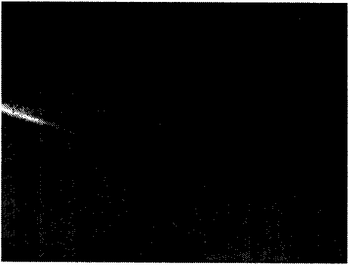
[0042]Put 200mg of chitosan in a 10ml small beaker, add 9ml of 0.1% acetic acid, after the chitosan is completely dissolved, sterilize by high pressure steam (121°C, 20min), put 5g of β-sodium glycerophosphate in a 10ml small beaker, add 10ml of culture medium And 0.22μm filter membrane filtration sterilization, after 250mg hydroxyethyl cellulose irradiation sterilization, dissolve into 2.5% solution with 10ml culture medium, the three can be mixed according to the ratio of 4:1:1 to prepare neutral pH value, room temperature Liquid gel with good fluidity, it will be in the form of solid gel after being placed in the incubator for about 10 minutes ( figure 1 ).

Embodiment 2

[0043] Embodiment 2: Histocompatibility detection of chitosan hydrogel

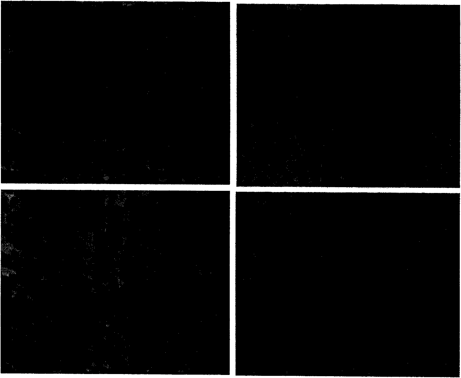
[0044] Chitosan hydrogel 0.1ml was injected and implanted into the thigh muscles of S-D rats, and they were killed at 1, 2, 3, and 4 weeks respectively, and 2 animals were taken each time. After the specimens were taken out, paraffin sections were taken, and HE staining was used to observe the inflammatory reaction Case. One week after operation, chitosan hydrogel was widely distributed in the myocardial tissue space, and a little inflammatory cells were mixed in between ( figure 2 A); 2 weeks after operation, there was obvious inflammatory cell infiltration around the chitosan hydrogel ( figure 2 B); 3 weeks after operation, no obvious chitosan hydrogel could be seen in the group, but the inflammatory cell infiltration was still obvious ( figure 2 C); 4 weeks after operation, no obvious chitosan hydrogel-like substance remained, but a small amount of inflammatory cell infiltration could still be see...

Embodiment 3

[0045] Example 3: Selection of Animals and Preparation of Myocardial Infarction

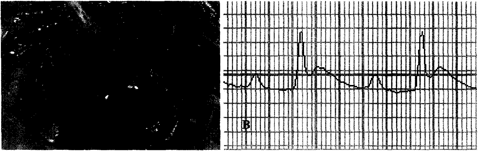
[0046] S-D rats (about 225-300g) were used in the experiment, sodium pentobarbital (40mg / kg body weight) was used for intraperitoneal anesthesia, and connected to an animal microventilator. A thoracotomy was performed, and the pericardium was cut to fully expose the heart. At 2mm below the left atrial appendage, suture with 0-7 silk suture. After ligation, the left ventricular wall became pale and the wall motion was weakened ( image 3 A). Cardiac monitoring showed that the ST segment of leads I and II was significantly elevated, indicating that the animal model of coronary artery infarction was successfully prepared ( image 3 B).


PUM


Abstract

The invention discloses an injectable myocardial tissue engineering product based on temperature-sensitive chitosan aquagel. Concretely, the temperature-sensitive chitosan aquagel is used as a support material and carries one or a plurality of different blood vessel growth promotion factors, such as bFGF, PDGF-BB, VEGF, and the like, and the condition that the temperature-sensitive chitosan aquagel restores a myocardial infarction area is observed after the temperature-sensitive chitosan aquagel is injected and transplanted to the specific area of an animal myocardial infarction model. The temperature-sensitive myocardial tissue engineering product established by applying the support material can promote the growth of myocardial capillary vessels and arteriolae, can increase the growth of sarcogenic fibres, can promote the regeneration of myocardial tissues, can increase the locular wall thickness of the infarction area, can remodel the shape of an original ventricle and can improve the function of the heart. The invention has simple operation technology and mild enforcement condition, provides a new product for the myocardial tissue engineering and has an important meaning for the clinical development of treating heart diseases to tissue engineering cardiac muscles.

Description


Claims


Application Information

Patent Timeline
Owner INST OF BASIC MEDICAL SCI ACAD OF MILITARY MEDICAL SCI OF PLA
Why Eureka
Try Eureka